Clean FRCS notes with inline figures
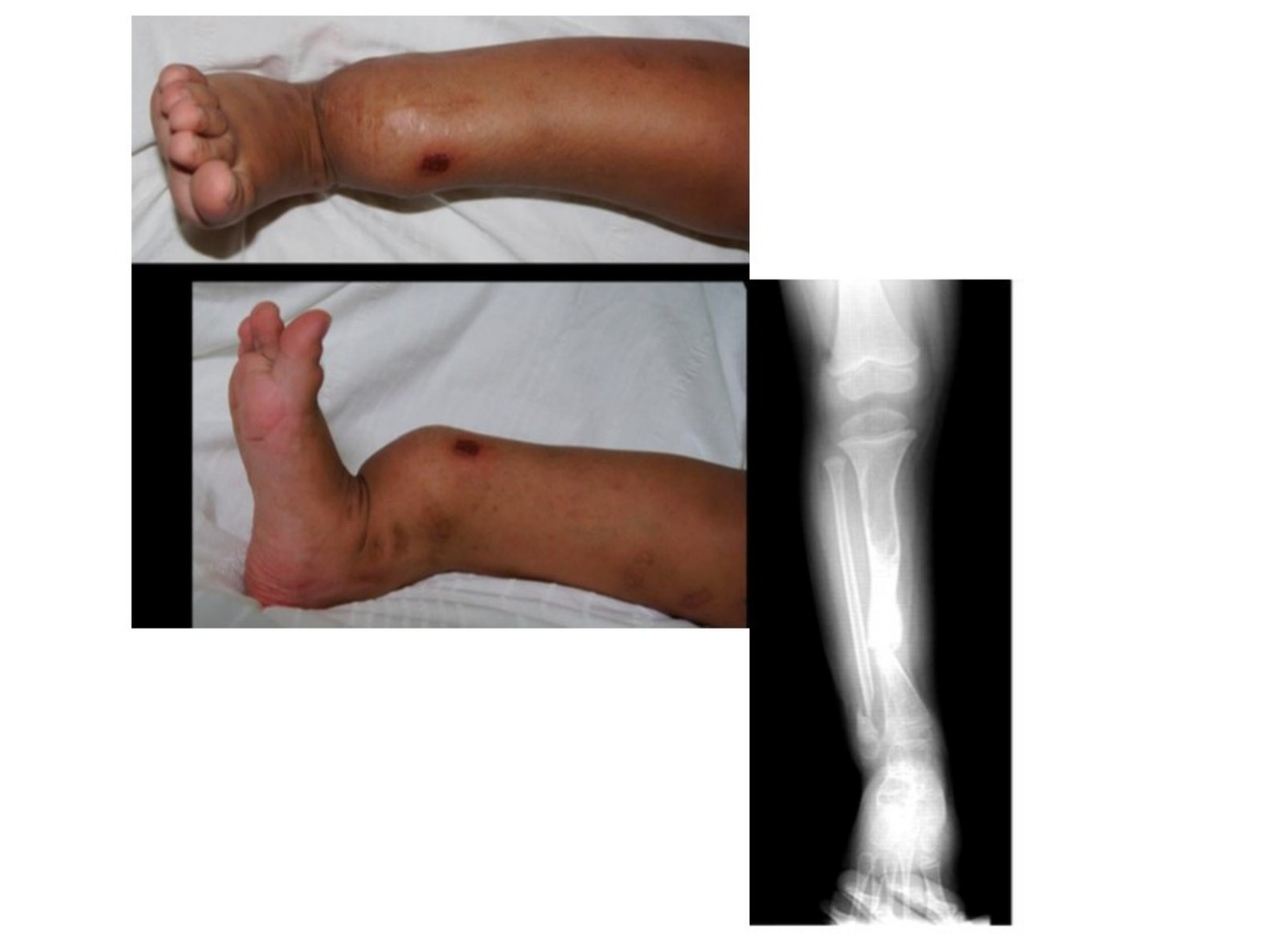
•Clinically photo showing patient’s right leg with abnormal angulation at level of distal tibia, with apex anterior, on frontal view there is no significant angulation (AL bowing), multiple hyperpigmented skin lesions ?scar/ café au lait spots
•Any neurofibroma / axillary freckles
•Anterior bowing of tibia is likely pathological
•Xray showing abnormalities of distal tibia and fibula, # with ?pseudoarthrosis over tibia, fibula length is normal → Dx Tibial pseudoarthrosis
Differential diagnosis
•NF with tibial pseudoarthrosis
•Tibial hemimelia (anterolateral bowing)
•Fibrous dysplasia
•Trauma with fracture callus
•Tumor (EWS, osteofibrous dysplasia, adamantinoma)
•Osteomyelitis
Crawford classification. Others: Anderson, Boyd
| Type / group | Definition / note |
|---|---|
| I | Dense cortex: no need brace, may never fracture. |
| II | Widened cortex, narrow IM + trabeculation failure: brace. union rate 91% |
| III | Cystic: pre fracture. Early fixation. Union rate 75% |
| IV | Dysplastic, fracture, cyst, pseudoarthrosis. Union rate 70% |
•Hx, PE
•Age, FHx, associated abnormalities (of NF and tibial hemimelia), previous treatment, LLD, angular deformity, joint stiffness/ OA
General management aim:
•Promote union → Prevent fracture
•Correct deformity + Equal leg length
•Natural history: vicious cycle of limb angulation, repeated #, LLD, joint stiffness
•Check/perform counsel patient’s family from the outset about possibility of amputation
•If prefracture – Clampshell brace, no evidence to suggest bracing will prevent fracture or deformity
•If impending fracture/ fracture / pseudoarthrosis, MRI first to see extent of pseudoarthrosis and how much to resect
•4 in 1 osteosynthesis
•Resection of pseudoarthrosis + fibrotic periosteum
•Correct angular deformity → 3 options: bone grafting, acute shortening, bone transport
•Enhance biology with BMP2,7, periosteal flap, autogenous bone graft (limited source from child) [E: iliac graft]
•Stable fixation fixation by ilizarov + IMN (through ankle joint) [often most difficult part due to small bone → William’s rod]
•Serial observation + bracing until maturity
•EPOS (european paediatric orthopaedic society) 2000 Gill
•340 patients
•Pre fracture clampshell brace
•# → ilizarov (provide compression, also allow lengthening/ bone transport) → IM nail (antegrade rush pin, or splint ankle to control distal fragment and to prevent future #, or William rod – through STJ and TTJ if <4yo, proximalise it if postop 2 years and not grown out of ankle joint yet )
•Poor prognostic factors are 1. young age, 2. advanced Crawford staging, 3. NF (not mentioned: fibular pseudoarthrosis)
•3 unsuccessful OT/ severe LLD (>15cm)/ non functional → amputate (SYMES, prosthesis protect pseudoarthrosis)
Argument for timing of surgery
•For older – difficult to provide stable fixation with any technique in small child, even with ilizarov. Also difficult to ask them to weight bear with the frame, which is needed to stimulate bone growth
•For younger – if delay till older. A braced and protected leg cannot develop normally and will become dysplastic, lose function, become shorter due to growth retardation.
•Therefore, based on EPOS – reasonable success between 3-6 yo (the fibromatosis of the tissue found in the area of pseudoarthrosis has greater osteolytic activity in smaller children)
Janus:
Principle mx:
•Prevent #
•Coz once #, diffi to heal
key point in OT:
•Cross union
•4 in 1 procedure
•Coz patho harmartoma/ periosteaum
•Periosteal graft from iliac crest
•Excise harmartoma, recanalize
•X guided growth – coz deformity is in diaphysis not in physis
•So osteotomy
MRI not necessary if XR definite craford 2
Intraop assessment x firbrous tissue/ harmartoma
•Functional outcome, at maturity
•FWB 40%
•LLD 60%
•Walk unlimited 70%
•N Ankle 30%
•Sports <30%
Pathology:
•Growth of fibrous harmatoma in pathological periosteum → strangulation of blood supply, decrease osteogenicity, increased osteoclastic resorption
•Dysplastic bone → failure of new bone formation and segmental osseous weakening
•Anterolateral bowing of tibia associated with NF (50%), ED syndrome, amnionic band syndrome
Why is Ilizarov the method of choice?
According to EPOS study, advantage:
•Excellent stability
•Allows complete resection of the pseudoarthritic area regardless of the amount of resected segment because this device allows lengthening or segmental bone transport
•Enables weight bearing , which stimulates healing of bone and soft tissue
•Can also be used to transport the fibula distally, avoid valgus ankle deformity, which is a problem commonly overlooked and underestimated
•Bone transport vs acute shortening + LLD correction via proximal metaphyseal lengthening after corticotomy
•Bone transport lower fusion rate
•Deep muscles and periosteum move with the transported segment but the superficial muscle and fascia move very little
•During transport, major vessels lengthen in the region of regeneration and become tortuous distally, causing kinking of vessels
•→ EPOS study suggest acute shortening + LLD correction via proximal metaphyseal lengthening after corticotomy