FRCS Paediatric Ortho — improved notes
FRCS Paediatric Ortho — visual inline notes

058. Rickets / osteomalacia

visual-inline rebuild2 extracted figuresspeaker notes XML61 study lines3 Q&A

Individual slide images have been cropped from the rendered slide and inserted beside the matching note section. The full slide remains below only for layout/audit.

Clean FRCS notes with inline figures

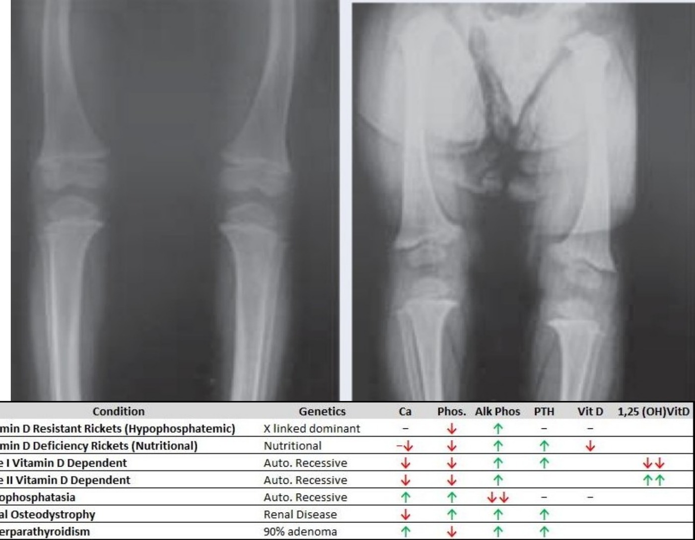
Xray LL of skeletally immature patient
Radiograph / imaging
Fig 1. Radiograph / imagingtop left
Classification / diagram
Fig 2. Classification / diagrambottom left
•Increased physeal width in general, cupping of metaphyseal area
•Bowing of bilateral tibia
•No coxa vara
•Systemic disease affecting physis
•Ddx: Rickets, MED, SED, achondroplasia

History:

Birth and family history

•Diet

Examination

•Hyperlaxity
•Generalized weakness
•Deformity
•Rickets features
•Syndromic features
•Ricket / Osteomalacia
•Defect in mineralization of osteoid matrix and chondroid (in zone of calcification) caused by inadequate calcium and phosphate
•Ca / PO4 homeostasis problem → impaired calcification of cartilage matrix of growing long bones → leads to increased physeal width and cortical thinning and bowing

Clinical:

•Short stature (proportionate)
•Frontal bossing + caput quadratum
•Craniotabes (softening of skull)
•Dolichocephaly (skull long posterior but thin width, due to premature closure of sutures)
•Pigeon chest
•Rachitic rosary
•Harrison’ sulcus
•Cat back kyphosis
•LL alignment abnormalities (SCFE, genu varum)
•Prominent distal wrist epiphysis

Biochemical:

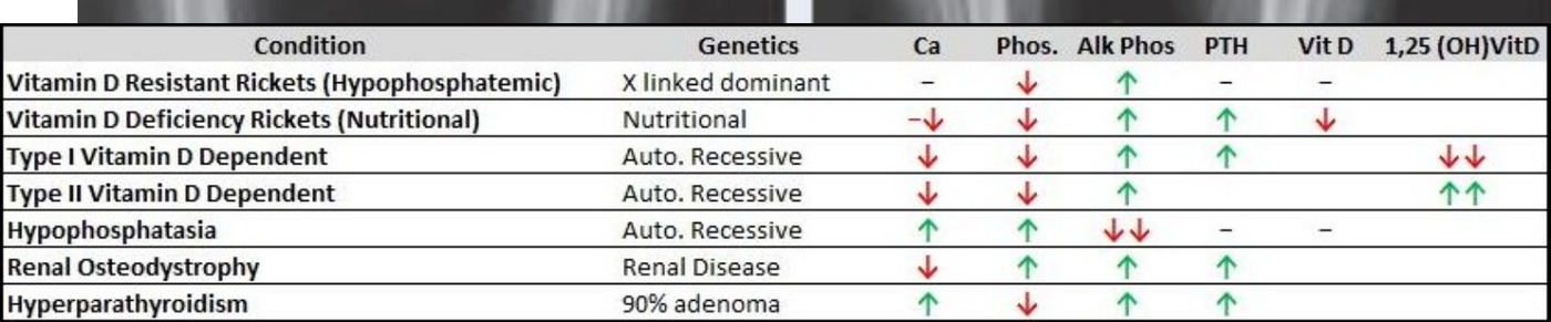
•Decreased Ca, PO4
•Increased ALP and PTH (except in Familial hypophosphatemic rickets and hypophosphatasia)
•Vit D level depends on cause
•(see chart above)

Xray:

•Long bone bowing
•Looser’s zone / pseudofracture (transverse radiolucency due to unmineralized osteoid seam) → weight bearing bone on compression side (insufficiency fracture), non weight bearing bone at nutrient foramen
•Metaphysis flaring in paintbrush (indistinct) and cupping (egg in a cup)
•Widened physis (at zone of hypertrophy)
•+/- osteopenia
•Cod-fish vertebrae
•(Hypophosphatemia: bowing more over diaphysis)
•Histology: unmineralized osteoid seam
•Causes
•Intake: nutritional problem, lack of sunlight exposure, GI problem

Processing:

•Vitamin D dependent rickets
Type / groupDefinition / note
Type Idefective 1α hydroxylase
Type IIend organ insensitivity to 1,25α hydroxylase

Recycling:

•Vitamin D resistant rickets (X linked dominant familial hypophosphatemic rickets) – PHEX (encode for endopeptidase cleaving FGF23), cannot chelate FGFR23, cannot reabsorb PO4 in the proximal tubules, cannot mineralize → more diaphyseal bowing rather than bowing a metaphysis (New drug, Burozumab)
•Hypophosphatasia – mutation in tissue nonspecific isoenzyme of ALP → reduced ALP, decreased synthesis of phosphate, decrease mineralization (increase urine phosphoethanol-amine)

Treatment

•Underlying cause
•Dietary input → Vit D3 supplement (5000IU/day)
•Vitamin dependent rickets
•Type 1 → physiologic doses (1-2 micrograms/day) of 1,25-(OH)2-Vit D (Calcitriol)
•Type 2 → high dose Vitamin D
•Hypophosphateamic syndrome → Calcitriol +/- phosphate (1g)
•Mild deformity→ watchful observation for remodeling
•Severe deformity affect ADL→ deformity correction

Direct Q&A

Q1 What are the radiographic findings for Rickets / osteomalacia?

Increased physeal width in general, cupping of metaphyseal area

•Bowing of bilateral tibia
•No coxa vara
•Systemic disease affecting physis
•Ddx: Rickets, MED, SED, achondroplasia
Q2 What examination findings should be assessed in Rickets / osteomalacia?

Hyperlaxity

•Generalized weakness
•Deformity
•Rickets features
•Syndromic features
•Ricket / Osteomalacia
•Defect in mineralization of osteoid matrix and chondroid (in zone of calcification) caused by inadequate calcium and phosphate
•Ca / PO4 homeostasis problem → impaired calcification of cartilage matrix of growing long bones → leads to increased physeal width and cortical thinning and bowing
Q3 How is Rickets / osteomalacia managed?

Underlying cause

•Dietary input → Vit D3 supplement (5000IU/day)
•Vitamin dependent rickets
•Type 1 → physiologic doses (1-2 micrograms/day) of 1,25-(OH)2-Vit D (Calcitriol)
•Type 2 → high dose Vitamin D
•Hypophosphateamic syndrome → Calcitriol +/- phosphate (1g)
•Mild deformity→ watchful observation for remodeling
•Severe deformity affect ADL→ deformity correction

Raw speaker-note hierarchy audit

#lvlmarLindentlogicaloriginal note line
10NoneNone0Xray LL of skeletally immature patient
20NoneNone0Increased physeal width in general, cupping of metaphyseal area
30NoneNone0Bowing of bilateral tibia
40NoneNone0No coxa vara
50NoneNone0Systemic disease affecting physis
60NoneNone0Ddx: Rickets, MED, SED, achondroplasia
70NoneNone0History:
80NoneNone0Birth and family history
90NoneNone0Diet
100NoneNone0PE:
110NoneNone0Hyperlaxity
120NoneNone0Generalized weakness
130NoneNone0Deformity
140NoneNone0Rickets features
150NoneNone0Syndromic features
160NoneNone0Ricket / Osteomalacia
170NoneNone0Defect in mineralization of osteoid matrix and chondroid (in zone of calcification) caused by inadequate calcium and phosphate
180NoneNone0Ca / PO4 homeostasis problem → impaired calcification of cartilage matrix of growing long bones → leads to increased physeal width and cortical thinning and bowing
190NoneNone0Clinical:
200171450-1714501Short stature (proportionate)
210171450-1714501Frontal bossing + caput quadratum
220171450-1714501Craniotabes (softening of skull)
230171450-1714501Dolichocephaly (skull long posterior but thin width, due to premature closure of sutures)
240171450-1714501Pigeon chest
250171450-1714501Rachitic rosary
260171450-1714501Harrison’ sulcus
270171450-1714501Cat back kyphosis
280171450-1714501LL alignment abnormalities (SCFE, genu varum)
290171450-1714501Prominent distal wrist epiphysis
300NoneNone0Biochemical:
310NoneNone0Decreased Ca, PO4
320NoneNone0Increased ALP and PTH (except in Familial hypophosphatemic rickets and hypophosphatasia)
330NoneNone0Vit D level depends on cause
340NoneNone0(see chart above)
350NoneNone0Xray:
360171450-1714501Long bone bowing
370171450-1714501Looser’s zone / pseudofracture (transverse radiolucency due to unmineralized osteoid seam) → weight bearing bone on compression side (insufficiency fracture), non weight bearing bone at nutrient foramen
380171450-1714501Metaphysis flaring in paintbrush (indistinct) and cupping (egg in a cup)
390171450-1714501Widened physis (at zone of hypertrophy)
400171450-1714501+/- osteopenia
410171450-1714501Cod-fish vertebrae
420NoneNone0(Hypophosphatemia: bowing more over diaphysis)
430NoneNone0Histology: unmineralized osteoid seam
440NoneNone0Causes
450171450-1714501Intake: nutritional problem, lack of sunlight exposure, GI problem
460171450-1714501Processing:
471628650-1714502Vitamin D dependent rickets
4821085850-1714503Type I: defective 1α hydroxylase
4921085850-1714503Type II: end organ insensitivity to 1,25α hydroxylase
500171450-1714501Recycling:
511628650-1714502Vitamin D resistant rickets (X linked dominant familial hypophosphatemic rickets) – PHEX (encode for endopeptidase cleaving FGF23), cannot chelate FGFR23, cannot reabsorb PO4 in the proximal tubules, cannot mineralize → more diaphyseal bowing rather than bowing a metaphysis (New drug, Burozumab)
521628650-1714502Hypophosphatasia – mutation in tissue nonspecific isoenzyme of ALP → reduced ALP, decreased synthesis of phosphate, decrease mineralization (increase urine phosphoethanol-amine)
530NoneNone0Treatment
540NoneNone0Underlying cause
550171450-1714501Dietary input → Vit D3 supplement (5000IU/day)
560171450-1714501Vitamin dependent rickets
571628650-1714502Type 1 → physiologic doses (1-2 micrograms/day) of 1,25-(OH)2-Vit D (Calcitriol)
581628650-1714502Type 2 → high dose Vitamin D
590171450-1714501Hypophosphateamic syndrome → Calcitriol +/- phosphate (1g)
600NoneNone0Mild deformity→ watchful observation for remodeling
610NoneNone0Severe deformity affect ADL→ deformity correction

Improved semantic hierarchy used for the study notes

#semantic levelsection?rewritten line
10yesXray LL of skeletally immature patient
21Increased physeal width in general, cupping of metaphyseal area
31Bowing of bilateral tibia
41No coxa vara
51Systemic disease affecting physis
61Ddx: Rickets, MED, SED, achondroplasia
70yesHistory:
80yesBirth and family history
91Diet
100yesExamination
111Hyperlaxity
121Generalized weakness
131Deformity
141Rickets features
151Syndromic features
161Ricket / Osteomalacia
171Defect in mineralization of osteoid matrix and chondroid (in zone of calcification) caused by inadequate calcium and phosphate
181Ca / PO4 homeostasis problem → impaired calcification of cartilage matrix of growing long bones → leads to increased physeal width and cortical thinning and bowing
190yesClinical:
201Short stature (proportionate)
211Frontal bossing + caput quadratum
221Craniotabes (softening of skull)
231Dolichocephaly (skull long posterior but thin width, due to premature closure of sutures)
241Pigeon chest
251Rachitic rosary
261Harrison’ sulcus
271Cat back kyphosis
281LL alignment abnormalities (SCFE, genu varum)
291Prominent distal wrist epiphysis
300yesBiochemical:
311Decreased Ca, PO4
321Increased ALP and PTH (except in Familial hypophosphatemic rickets and hypophosphatasia)
331Vit D level depends on cause
341(see chart above)
350yesXray:
361Long bone bowing
371Looser’s zone / pseudofracture (transverse radiolucency due to unmineralized osteoid seam) → weight bearing bone on compression side (insufficiency fracture), non weight bearing bone at nutrient foramen
381Metaphysis flaring in paintbrush (indistinct) and cupping (egg in a cup)
391Widened physis (at zone of hypertrophy)
401+/- osteopenia
411Cod-fish vertebrae
421(Hypophosphatemia: bowing more over diaphysis)
431Histology: unmineralized osteoid seam
441Causes
451Intake: nutritional problem, lack of sunlight exposure, GI problem
460yesProcessing:
472Vitamin D dependent rickets
483Type I: defective 1α hydroxylase
493Type II: end organ insensitivity to 1,25α hydroxylase
500yesRecycling:
512Vitamin D resistant rickets (X linked dominant familial hypophosphatemic rickets) – PHEX (encode for endopeptidase cleaving FGF23), cannot chelate FGFR23, cannot reabsorb PO4 in the proximal tubules, cannot mineralize → more diaphyseal bowing rather than bowing a metaphysis (New drug, Burozumab)
522Hypophosphatasia – mutation in tissue nonspecific isoenzyme of ALP → reduced ALP, decreased synthesis of phosphate, decrease mineralization (increase urine phosphoethanol-amine)
530yesTreatment
541Underlying cause
551Dietary input → Vit D3 supplement (5000IU/day)
561Vitamin dependent rickets
572Type 1 → physiologic doses (1-2 micrograms/day) of 1,25-(OH)2-Vit D (Calcitriol)
582Type 2 → high dose Vitamin D
591Hypophosphateamic syndrome → Calcitriol +/- phosphate (1g)
601Mild deformity→ watchful observation for remodeling
611Severe deformity affect ADL→ deformity correction