Clean FRCS notes with inline figures
Achondroplasia
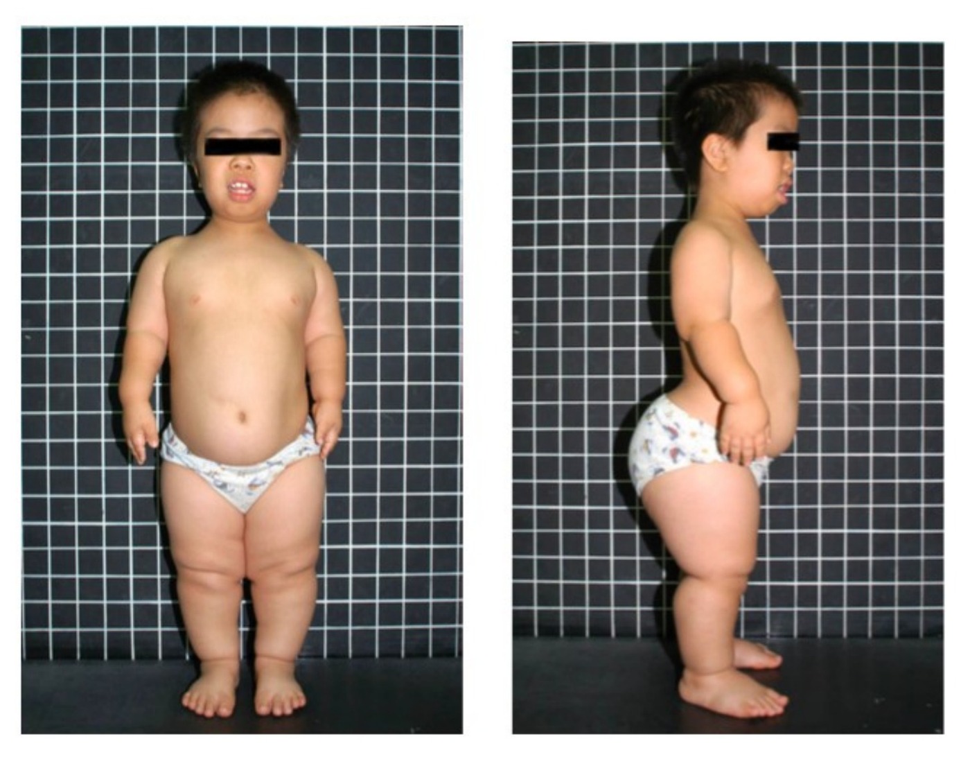
•Clinical photo showing patient of short stature which is disproportional (as patient’s hand can only reach pelvis instead of thigh in usual population). Head also appears large
•It is rhizomelic short stature as patient’s humerus appears shorter than his forearm and thigh similar length as leg (usually 0.8)
•Dysmorphism: frontal bossing, hypoplastic midface, otitis media, normal IQ
•Limb: elbow flexion contracture (posterior RH dislocation), trident hand, Genu varum
•Trunk: TL kyphosis, LS hyperlordosis (spinal stenosis due to short pedicle) (cervical myelopathy due to foramen magnum stenosis). Protruberant abdomen. Flat chest
Further PE:
•1. Standing and seating height (standing <3rd percentile, sitting normal)
•2. Measure limb length (rhizomelic shortening)
•3. Ligamentous laxity
•4. Check elbow ROM, posterior radial head dislocation
•Ddx (disproportionate short limb dwarfism)
•Hypochondroplasia
•PseudoAchondroplasia
•Leri Wiel Dyschondrosteosis
•MED
Rubin classification: physeal hypoplasia
•Most common cause of disproportionate dwarfism
•Genetics: AD; Sporadic mutation > 80% (risks x advanced paternal age), caused by mutation of FGFR3 on chromosome 4 (glycine base being substituted by an arginine base → prolonged receptor activation after ligand binding and result in excessive growth limitation)
•Mutation leads to abnormal chondroid production by chondroblasts in the proliferative zone during enchondral bone formation at the physis (IM ossification not affected)
•Quantitative cartilage defect
Diagnosis: prenatal USG
Other ortho problem:
•Foramen magnum stenosis
•Spinal stenosis
•Radial head subluxation, elbow FFC
•Genu varum
Difficulties in spinal surgery:
•Positioning
•Exposure – increase lumbar lordosis