FRCS Paediatric Ortho — improved notes
FRCS Paediatric Ortho — visual inline notes

094. Spine embryology

visual-inline rebuild3 extracted figuresspeaker notes XML10 study lines1 Q&A

Individual slide images have been cropped from the rendered slide and inserted beside the matching note section. The full slide remains below only for layout/audit.

Clean FRCS notes with inline figures

Spine embryology
Radiograph / imaging
Fig 1. Radiograph / imagingtop left
Radiograph / imaging
Fig 2. Radiograph / imagingmiddle centre
Radiograph / imaging
Fig 3. Radiograph / imagingtop centre
•3 embryological layers: ectoderm, mesoderm, endoderm
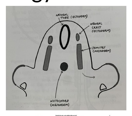
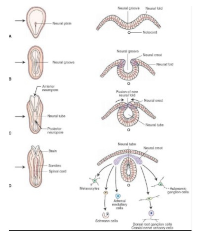
•At 3 weeks, they aggregate at midline, the ectoderm infolds onto itself in the midline and form the neural tube, which is the precursor for CNS development. Dorsal to neurotube are the neural crest cells, which become the PNS. The mesoderm forms the notochord, which induces the process of neurylation and becomes the nucleus pulposus at maturity.
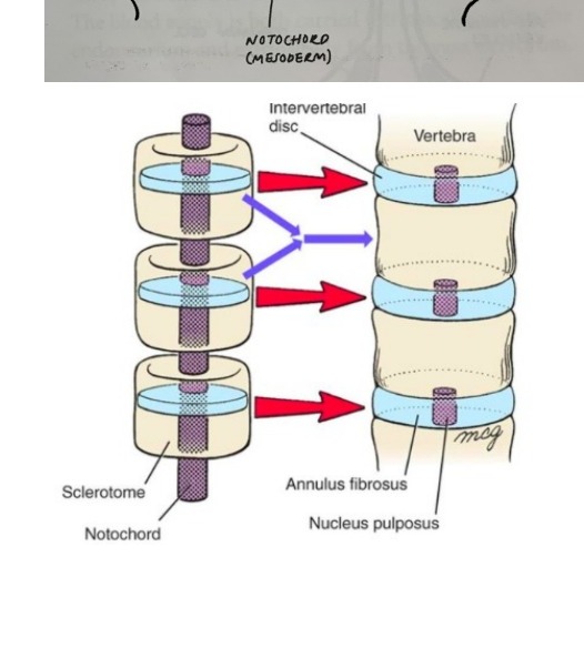
•At 4 weeks, paraxial mesoderm also aggregate in the midline to form somites (dermatome, myotome and sclerotome), which unite around the notochord to form the vertebral body and around the neutral tube to form the neural arch. Each somite forms one disc plus the adjoining cranial and caudal halves of the vertebra. The central part undergoes apoptosis to form the disc. And the superior and inferior parts fuse to form a vertebral body (von Eber fissure)
•At 6 weeks, the spine undergoes chondrification
•At 9 weeks, the primary ossification center is formed, consisting of 1 primary at body, and 2 primary at neural arch
•At 2-6 years old, the arches fuse with the body
•At 12 years old, the secondary ossification centers appear at the spinous process, transverse process, apophyseal rings
•At 25 years old, the secondary fuses to the primary ossification center
•Apophyseal ring avulsion fracture can be associated with PID in teenager

Direct Q&A

Q1 What are the diagnostic findings and management points for Spine embryology?

Spine embryology

•3 embryological layers: ectoderm, mesoderm, endoderm
•At 3 weeks, they aggregate at midline, the ectoderm infolds onto itself in the midline and form the neural tube, which is the precursor for CNS development. Dorsal to neurotube are the neural crest cells, which become the PNS. The mesoderm forms the notochord, which induces the process of neurylation and becomes the nucleus pulposus at maturity.
•At 4 weeks, paraxial mesoderm also aggregate in the midline to form somites (dermatome, myotome and sclerotome), which unite around the notochord to form the vertebral body and around the neutral tube to form the neural arch. Each somite forms one disc plus the adjoining cranial and caudal halves of the vertebra. The central part undergoes apoptosis to form the disc. And the superior and inferior parts fuse to form a vertebral body (von Eber fissure)
•At 6 weeks, the spine undergoes chondrification
•At 9 weeks, the primary ossification center is formed, consisting of 1 primary at body, and 2 primary at neural arch
•At 2-6 years old, the arches fuse with the body
•At 12 years old, the secondary ossification centers appear at the spinous process, transverse process, apophyseal rings
•At 25 years old, the secondary fuses to the primary ossification center
•Apophyseal ring avulsion fracture can be associated with PID in teenager

Raw speaker-note hierarchy audit

#lvlmarLindentlogicaloriginal note line
10NoneNone0Spine embryology
20NoneNone03 embryological layers: ectoderm, mesoderm, endoderm
30NoneNone0At 3 weeks, they aggregate at midline, the ectoderm infolds onto itself in the midline and form the neural tube, which is the precursor for CNS development. Dorsal to neurotube are the neural crest cells, which become the PNS. The mesoderm forms the notochord, which induces the process of neurylation and becomes the nucleus pulposus at maturity.
40NoneNone0At 4 weeks, paraxial mesoderm also aggregate in the midline to form somites (dermatome, myotome and sclerotome), which unite around the notochord to form the vertebral body and around the neutral tube to form the neural arch. Each somite forms one disc plus the adjoining cranial and caudal halves of the vertebra. The central part undergoes apoptosis to form the disc. And the superior and inferior parts fuse to form a vertebral body (von Eber fissure)
50NoneNone0At 6 weeks, the spine undergoes chondrification
60NoneNone0At 9 weeks, the primary ossification center is formed, consisting of 1 primary at body, and 2 primary at neural arch
70NoneNone0At 2-6 years old, the arches fuse with the body
80NoneNone0At 12 years old, the secondary ossification centers appear at the spinous process, transverse process, apophyseal rings
90NoneNone0At 25 years old, the secondary fuses to the primary ossification center
100NoneNone0Apophyseal ring avulsion fracture can be associated with PID in teenager

Improved semantic hierarchy used for the study notes

#semantic levelsection?rewritten line
10Spine embryology
213 embryological layers: ectoderm, mesoderm, endoderm
31At 3 weeks, they aggregate at midline, the ectoderm infolds onto itself in the midline and form the neural tube, which is the precursor for CNS development. Dorsal to neurotube are the neural crest cells, which become the PNS. The mesoderm forms the notochord, which induces the process of neurylation and becomes the nucleus pulposus at maturity.
41At 4 weeks, paraxial mesoderm also aggregate in the midline to form somites (dermatome, myotome and sclerotome), which unite around the notochord to form the vertebral body and around the neutral tube to form the neural arch. Each somite forms one disc plus the adjoining cranial and caudal halves of the vertebra. The central part undergoes apoptosis to form the disc. And the superior and inferior parts fuse to form a vertebral body (von Eber fissure)
51At 6 weeks, the spine undergoes chondrification
61At 9 weeks, the primary ossification center is formed, consisting of 1 primary at body, and 2 primary at neural arch
71At 2-6 years old, the arches fuse with the body
81At 12 years old, the secondary ossification centers appear at the spinous process, transverse process, apophyseal rings
91At 25 years old, the secondary fuses to the primary ossification center
101Apophyseal ring avulsion fracture can be associated with PID in teenager