Clean FRCS notes with inline figures
| Type / group | Definition / note |
|---|---|
| X | ray of skeletally immature patient |
•Bilateral elbow
•On the Xray on top the relationship of the proximal ulna and radius are maintained, but the relationship of the humerus and the ulna & radius is disrupted with the later displaced posteromedially
•This is likely a transphyseal fracture of distal humerus (more likely than elbow dislocation as physis is biomechanically the weakest location in distal humerus)
•1. Normal alignment between the proximal radius and the capitellum (radiocapitellar line)
•2. Normal alignment of the proximal radius and ulna with each other
•Vs elbow dislocation: PM displacement in transphyseal injury
•Vs lateral condyle fracture: lateral condyle radial head relationship
•Vs low SCH fracture: wider bone contact in transphyeal, less displacement
•Hyperextension injury (birth injury/ abuse/ trauma, usu <2y)
Anatomy:
•Medial epicondyle involved at young age
•Physis moves distally as one grow, V shaped cleft
•Relationship of radius and capitellum is preserved
•Large fragment, usually less rotation and tilting
•Blood supply good, but medial crista of trochlea blood supply may be disrupted → cubital varus
DeLee classification (this type of fracture almost only < 7y)
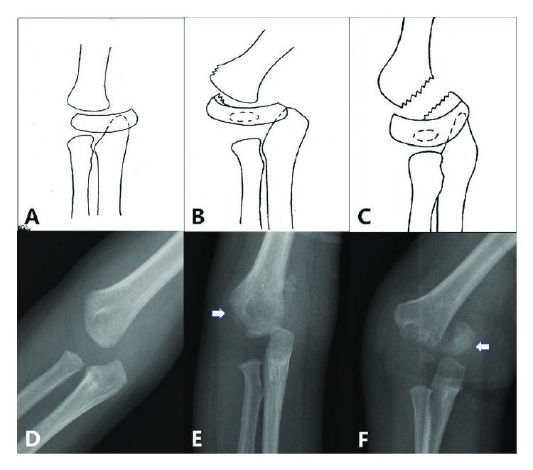
| Type / group | Definition / note |
|---|---|
| Type A | infant-7m. No lateral condyle ossification center (SH 1) |
| Type B | 7m-3y. Lateral condyle ossified (SH 1, 2) |
| Type C | 3y – 7. large metaphyseal fragment exiting laterally → CR +/- OR + KWF + cast in pronation x 3/52 (+/- arthrogram) |
•Highly suspicious of NAI!!
Further Mx:
•MRI
•Management depends on timing of injury, displacement and DeLee type
•Acute undisplaced → CR + Cast in pronation x 3/52
•Acute displaced → CR +/- OR+ KWF + cast in pronation x 3/52
•Late (> 1/52) + displaced → DO NOT manipulate