FRCS Paediatric Ortho — improved notes
FRCS Paediatric Ortho — visual inline notes

013. SCFE/SUFE

visual-inline rebuild1 extracted figuresspeaker notes XML99 study lines8 Q&A

Individual slide images have been cropped from the rendered slide and inserted beside the matching note section. The full slide remains below only for layout/audit.

Clean FRCS notes with inline figures

Child complain of knee pain**

•“Medial knee pain in child with normal knee XR”
•SCREEN FOR HIP!!!
•Xray showing pelvis of skeletally immature patient with abnormalities over left proximal femur (looks to be around 12 yo based on GT, triradiate cartilage and LT)
•Slipped capital femoral epiphysis SCFE
•Klein’s line (Trethowan's Sign = when Klein line does not cut the epiphysis)
•Widened physis and decreased epiphyseal height
•Increased distance between the teardrop and the femoral neck metaphysis.
•Metaphyseal blanch sign of steel
•Carpener’s sign (normal AP: posterior acetabular margin cuts across the medial corner of the upper femoral metaphysis. In SUFE, the entire metaphysis is lateral to the posterior acetabular margin)

Signs of chronic SCFE:

•metaphysis smooth and sclerotic with callus formation
•metaphysis widens (coxa magna)
•No features of slip on the contralateral side (20% bilateral)

Differential is #

History

•1. Timing (chronicity)
•2. Able to walk at all (stability)
•3. Any preexisting illness or risk factors (endocrinopathy - hypothyroid, GH def, hypogonadism, renal failure)
•4. Sx on other side (20% bilateral, 80% chronic)

Examination

•See if patient can stand
•Position of limb
•Chronic – obligatory ER with flexion (Drenman’s sign)

Classification: chronicity, stability, severity

•Chronicity: pre-slip, acute, chronic, acute on chronic (no bearing on prognosis)
•Stability: Loder
•Stable <10% AVN
•Unstable 50% AVN
•Severity: Southwick angle in frog view, Wilson grade in AP view

Xray frog if stable, cross table if unstable
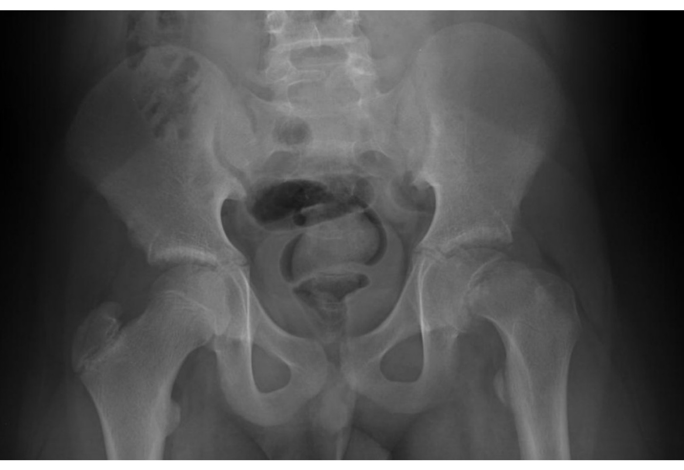
Radiograph / imaging - SCFE radiograph
Fig 1. Radiograph / imaging - SCFE radiographtop left

For Southwick classification (line perpendicular to base of epiphysis and line along shaft, compare with contralateral side)

•<30, 30-50, >50
•if bilateral slip, then Southwick angle is calculated by subtracting 12 degrees (normal angle)

Wilson’s grades: Epiphyseal-metaphyseal method (AP View)

•<1/3, 1/3-2/3, >2/3

Management

•Aim: to prevent further slip, promote physeal closure, prevent complications
•Natural history (Weinstein CORR 1996) – 28 untreated patients → increase displacement, degree of displacement is proportional to the risk of OA
•Unstable - O&T emergency, aim at preventing further slippage and allow physis to fuse in 6hrs (8-24hr)
•Go for in-situ pinning
•Stable, still pin in-situ, just not that urgent
•Also say whether go for prophylactic pin
•Risk factors for bilateral: Young (E: <10y), obese (E: not absolute), endocrinopathy (YOE)
Type / groupDefinition / note
EInability to come back fu / tell pain
•1. GA
•2. Xray guidance
•3. Supine on traction table
•4. NO traction / forceful manipulation / only serendipitous reduction
•5. Very careful during positioning
•6. Triangulation technique to define entry point. Expect anterior entry (anterior aspect of femoral neck at head neck junction)
•7. One pin aiming at center center position, crossing perpendicular to physis, with adequate purchase and tip below subchondral bone, cross physis by at least 4 threads
•8. Cannulated, titanium, 6.5mm partially threaded screw with reverse cutting flutes
•9. Avoid multiple puncture → chondrolysis
•10. Capsulotomy (theoretical benefit but no strong evidence to support)
•JPO Dodwell metanalysis 2019- no stat significant decrease in odds of AVN. But recommend routine capsulotomy for all unstable SCFE as it adds little to the surgical procedure and may minimise risk of devastating insult to femoral head
•Consider contralateral pinning if
•(22% contralateral slip in 18 month)
•Young <10y (open triradiate cartilage)
•Systemic disease (hypothyroid, GH def, hypogonadism, renal failure)
•Obese / retroverted neck
•?Lost to FU
•Radiological markers to aid decision making – posterior slope angle (>14deg – 83% risk) and modified oxford bone age

One pin vs two pin:

•Theoretically 2 pins more stable, BUT
•Check/perform avoid superior quadrant of femoral head (where retinacular artery enters)
•Posterior aspect also avoid (in-out-in can damage posterior blood supply)
•Grade 3 slips or above → 25% contact only, limited corridor for pins
•Karol JPO 1992: Resistance to further slip was not proportional to screw number. Single screw fixation in slipped epiphyses is recommended because the small gains in stiffness with a second screw do not offset the risk of complications.

Any other option for severe type:

•Refer to specialist center for consideration
•Open reduction and fixation with Modified Dunn’s procedure
•Based on anatomy that MFCA is protected by obturator externus muscles
•Incision like posterior approach, Z shaped incision to capsule, avoid obturator externus, anterior dislocation of hip
•Reduction and fix with multiple pins

Rehab:

•Stable – weight bear
•Unstable – NWB

Follow-up checklist

•Pathological side fixation, complication (early: recurrent slippage, chondrolysis, Late: AVN, OA, FAI)
•Contralateral side
•FU until epiphyseal closure
•What if patient p/w pain + stiffness in FU
•R/O infection

Xray to see joint space. Chondrolysis if <3mm

•RF: manipulation, immobilization, hardware through head
•Rx: supportive, ?arthrodesis

•Definition: Disorder of the proximal femoral physis that leads to slippage of the metaphysis relative to the femoral head
•SCFE is misnomer: femoral head does not move, metaphysis slip and rotates
•Chronicity: acute <3w, chronic >3w, acute on chronic >3w with more pain → no implication on prognosis, management
•Timing of operation and pinning of contralateral hip is BOTH CONTROVERSIAL
•May present with knee pain due to radiation by obturator nerve
•Deformity: Extension, ER, varus
•Loss flexion, IR, abduction
•Cam type FAI (anterior impingement)
•Accelerated osteoarthritis due to anterior impingement
•PIS vs surgical dislocation for severe slip
•Metanalysis 2017 J O&T: surgical dislocation 3% AVN but better patient satisfaction rates whereas PIS has 1.5% AVN rate and lower satisfaction rates

Direct Q&A

Q1 What signs or features are listed for SCFE/SUFE?

metaphysis smooth and sclerotic with callus formation

•metaphysis widens (coxa magna)
•No features of slip on the contralateral side (20% bilateral)
Q2 What history should be elicited for SCFE/SUFE?
•1. Timing (chronicity)
•2. Able to walk at all (stability)
•3. Any preexisting illness or risk factors (endocrinopathy - hypothyroid, GH def, hypogonadism, renal failure)
•4. Sx on other side (20% bilateral, 80% chronic)
Q3 What examination findings should be assessed in SCFE/SUFE?

See if patient can stand

•Position of limb
•Chronic – obligatory ER with flexion (Drenman’s sign)
Q4 How is SCFE/SUFE classified here?
•Chronicity: pre-slip, acute, chronic, acute on chronic (no bearing on prognosis)
•Stability: Loder
•Stable <10% AVN
•Unstable 50% AVN
•Severity: Southwick angle in frog view, Wilson grade in AP view
Q5 How is SCFE/SUFE managed?

Aim: to prevent further slip, promote physeal closure, prevent complications

•Natural history (Weinstein CORR 1996) – 28 untreated patients → increase displacement, degree of displacement is proportional to the risk of OA
•Unstable - O&T emergency, aim at preventing further slippage and allow physis to fuse in 6hrs (8-24hr)
•Go for in-situ pinning
•Stable, still pin in-situ, just not that urgent
•Also say whether go for prophylactic pin
•Risk factors for bilateral: Young (E: <10y), obese (E: not absolute), endocrinopathy (YOE)
•E:Inability to come back fu / tell pain
•1. GA
•2. Xray guidance
•3. Supine on traction table
•4. NO traction / forceful manipulation / only serendipitous reduction
•5. Very careful during positioning
•6. Triangulation technique to define entry point. Expect anterior entry (anterior aspect of femoral neck at head neck junction)
•7. One pin aiming at center center position, crossing perpendicular to physis, with adequate purchase and tip below subchondral bone, cross physis by at least 4 threads
•8. Cannulated, titanium, 6.5mm partially threaded screw with reverse cutting flutes
•9. Avoid multiple puncture → chondrolysis
•10. Capsulotomy (theoretical benefit but no strong evidence to support)
•JPO Dodwell metanalysis 2019- no stat significant decrease in odds of AVN. But recommend routine capsulotomy for all unstable SCFE as it adds little to the surgical procedure and may minimise risk of devastating insult to femoral head
•Consider contralateral pinning if
•(22% contralateral slip in 18 month)
•Young <10y (open triradiate cartilage)
•Systemic disease (hypothyroid, GH def, hypogonadism, renal failure)
•Obese / retroverted neck
•?Lost to FU
•Radiological markers to aid decision making – posterior slope angle (>14deg – 83% risk) and modified oxford bone age
Q6 What rehabilitation plan is listed for SCFE/SUFE?

Stable – weight bear

•Unstable – NWB
Q7 What should be checked on follow-up for SCFE/SUFE?

Pathological side fixation, complication (early: recurrent slippage, chondrolysis, Late: AVN, OA, FAI)

•Contralateral side
•FU until epiphyseal closure
•What if patient p/w pain + stiffness in FU
•R/O infection
Q8 What are the radiographic findings for SCFE/SUFE?

RF: manipulation, immobilization, hardware through head

•Rx: supportive, ?arthrodesis

Raw speaker-note hierarchy audit

#lvlmarLindentlogicaloriginal note line
10NoneNone0Child complain of knee pain**
20NoneNone0“Medial knee pain in child with normal knee XR”
30NoneNone0SCREEN FOR HIP!!!
40NoneNone0Xray showing pelvis of skeletally immature patient with abnormalities over left proximal femur (looks to be around 12 yo based on GT, triradiate cartilage and LT)
50NoneNone0Slipped capital femoral epiphysis SCFE
60228600-2286001Klein’s line (Trethowan's Sign = when Klein line does not cut the epiphysis)
70228600-2286001Widened physis and decreased epiphyseal height
80228600-2286001Increased distance between the teardrop and the femoral neck metaphysis.
90228600-2286001Metaphyseal blanch sign of steel
100228600-2286001Carpener’s sign (normal AP: posterior acetabular margin cuts across the medial corner of the upper femoral metaphysis. In SUFE, the entire metaphysis is lateral to the posterior acetabular margin)
110000Signs of chronic SCFE:
120171450-1714501metaphysis smooth and sclerotic with callus formation
130171450-1714501metaphysis widens (coxa magna)
140NoneNone0No features of slip on the contralateral side (20% bilateral)
150NoneNone0Differential is #
160NoneNone0Hx:
170NoneNone01. Timing (chronicity)
180NoneNone02. Able to walk at all (stability)
190NoneNone03. Any preexisting illness or risk factors (endocrinopathy - hypothyroid, GH def, hypogonadism, renal failure)
200NoneNone04. Sx on other side (20% bilateral, 80% chronic)
210NoneNone0PE:
220NoneNone0See if patient can stand
230NoneNone0Position of limb
240NoneNone0Chronic – obligatory ER with flexion (Drenman’s sign)
250NoneNone0Classification: chronicity, stability, severity
261NoneNone1Chronicity: pre-slip, acute, chronic, acute on chronic (no bearing on prognosis)
27145720001Stability: Loder
28291440003Stable <10% AVN
29291440003Unstable 50% AVN
301NoneNone1Severity: Southwick angle in frog view, Wilson grade in AP view
310000Xray frog if stable, cross table if unstable
320000For Southwick classification (line perpendicular to base of epiphysis and line along shaft, compare with contralateral side)
33145720001<30, 30-50, >50
34145720001if bilateral slip, then Southwick angle is calculated by subtracting 12 degrees (normal angle)
350000Wilson’s grades: Epiphyseal-metaphyseal method (AP View)
36145720001<1/3, 1/3-2/3, >2/3
370000Mx:
380000Aim: to prevent further slip, promote physeal closure, prevent complications
390000Natural history (Weinstein CORR 1996) – 28 untreated patients → increase displacement, degree of displacement is proportional to the risk of OA
400000Unstable - O&T emergency, aim at preventing further slippage and allow physis to fuse in 6hrs (8-24hr)
410000Go for in-situ pinning
420000Stable, still pin in-situ, just not that urgent
430000Also say whether go for prophylactic pin
440000Risk factors for bilateral: Young (E: <10y), obese (E: not absolute), endocrinopathy (YOE)
450000E:Inability to come back fu / tell pain
4600001. GA
4700002. Xray guidance
4800003. Supine on traction table
4900004. NO traction / forceful manipulation / only serendipitous reduction
5000005. Very careful during positioning
5100006. Triangulation technique to define entry point. Expect anterior entry (anterior aspect of femoral neck at head neck junction)
5200007. One pin aiming at center center position, crossing perpendicular to physis, with adequate purchase and tip below subchondral bone, cross physis by at least 4 threads
5300008. Cannulated, titanium, 6.5mm partially threaded screw with reverse cutting flutes
5400009. Avoid multiple puncture → chondrolysis
55000010. Capsulotomy (theoretical benefit but no strong evidence to support)
56145720001JPO Dodwell metanalysis 2019- no stat significant decrease in odds of AVN. But recommend routine capsulotomy for all unstable SCFE as it adds little to the surgical procedure and may minimise risk of devastating insult to femoral head
570000Consider contralateral pinning if
580000(22% contralateral slip in 18 month)
591685800-2286002Young <10y (open triradiate cartilage)
601685800-2286002Systemic disease (hypothyroid, GH def, hypogonadism, renal failure)
611685800-2286002Obese / retroverted neck
621685800-2286002?Lost to FU
630000Radiological markers to aid decision making – posterior slope angle (>14deg – 83% risk) and modified oxford bone age
640000One pin vs two pin:
650171450-1714501Theoretically 2 pins more stable, BUT
660171450-1714501Need to avoid superior quadrant of femoral head (where retinacular artery enters)
670171450-1714501Posterior aspect also avoid (in-out-in can damage posterior blood supply)
680171450-1714501Grade 3 slips or above -> 25% contact only, limited corridor for pins
690171450-1714501Karol JPO 1992: Resistance to further slip was not proportional to screw number. Single screw fixation in slipped epiphyses is recommended because the small gains in stiffness with a second screw do not offset the risk of complications.
700000Any other option for severe type:
710000Refer to specialist center for consideration
720000Open reduction and fixation with Modified Dunn’s procedure
730000Based on anatomy that MFCA is protected by obturator externus muscles
740000Incision like posterior approach, Z shaped incision to capsule, avoid obturator externus, anterior dislocation of hip
750000Reduction and fix with multiple pins
760NoneNone0Rehab:
770NoneNone0Stable – weight bear
780000Unstable – NWB
790000What to look for in FU?
800228600-2286001Pathological side fixation, complication (early: recurrent slippage, chondrolysis, Late: AVN, OA, FAI)
810228600-2286001Contralateral side
820NoneNone0FU until epiphyseal closure
830NoneNone0What if patient p/w pain + stiffness in FU
840NoneNone0R/O infection
850NoneNone0Xray to see joint space. Chondrolysis if <3mm
860NoneNone0RF: manipulation, immobilization, hardware through head
870NoneNone0Rx: supportive, ?arthrodesis
880NoneNone0=================================================================
890000Definition: Disorder of the proximal femoral physis that leads to slippage of the metaphysis relative to the femoral head
900000SCFE is misnomer: femoral head does not move, metaphysis slip and rotates
910228600-2286001Chronicity: acute <3w, chronic >3w, acute on chronic >3w with more pain → no implication on prognosis, management
920228600-2286001Timing of operation and pinning of contralateral hip is BOTH CONTROVERSIAL
930228600-2286001May present with knee pain due to radiation by obturator nerve
940000Deformity: Extension, ER, varus
950000Loss flexion, IR, abduction
960000Cam type FAI (anterior impingement)
970000Accelerated osteoarthritis due to anterior impingement
980000PIS vs surgical dislocation for severe slip
99145720001Metanalysis 2017 J O&T: surgical dislocation 3% AVN but better patient satisfaction rates whereas PIS has 1.5% AVN rate and lower satisfaction rates

Improved semantic hierarchy used for the study notes

#semantic levelsection?rewritten line
10Child complain of knee pain**
21“Medial knee pain in child with normal knee XR”
31SCREEN FOR HIP!!!
41Xray showing pelvis of skeletally immature patient with abnormalities over left proximal femur (looks to be around 12 yo based on GT, triradiate cartilage and LT)
51Slipped capital femoral epiphysis SCFE
61Klein’s line (Trethowan's Sign = when Klein line does not cut the epiphysis)
71Widened physis and decreased epiphyseal height
81Increased distance between the teardrop and the femoral neck metaphysis.
91Metaphyseal blanch sign of steel
101Carpener’s sign (normal AP: posterior acetabular margin cuts across the medial corner of the upper femoral metaphysis. In SUFE, the entire metaphysis is lateral to the posterior acetabular margin)
110yesSigns of chronic SCFE:
121metaphysis smooth and sclerotic with callus formation
131metaphysis widens (coxa magna)
141No features of slip on the contralateral side (20% bilateral)
150yesDifferential is #
160yesHistory
1711. Timing (chronicity)
1812. Able to walk at all (stability)
1913. Any preexisting illness or risk factors (endocrinopathy - hypothyroid, GH def, hypogonadism, renal failure)
2014. Sx on other side (20% bilateral, 80% chronic)
210yesExamination
221See if patient can stand
231Position of limb
241Chronic – obligatory ER with flexion (Drenman’s sign)
250yesClassification: chronicity, stability, severity
261Chronicity: pre-slip, acute, chronic, acute on chronic (no bearing on prognosis)
271Stability: Loder
283Stable <10% AVN
293Unstable 50% AVN
301Severity: Southwick angle in frog view, Wilson grade in AP view
310yesXray frog if stable, cross table if unstable
320yesFor Southwick classification (line perpendicular to base of epiphysis and line along shaft, compare with contralateral side)
331<30, 30-50, >50
341if bilateral slip, then Southwick angle is calculated by subtracting 12 degrees (normal angle)
350yesWilson’s grades: Epiphyseal-metaphyseal method (AP View)
361<1/3, 1/3-2/3, >2/3
370yesManagement
381Aim: to prevent further slip, promote physeal closure, prevent complications
391Natural history (Weinstein CORR 1996) – 28 untreated patients → increase displacement, degree of displacement is proportional to the risk of OA
401Unstable - O&T emergency, aim at preventing further slippage and allow physis to fuse in 6hrs (8-24hr)
411Go for in-situ pinning
421Stable, still pin in-situ, just not that urgent
431Also say whether go for prophylactic pin
441Risk factors for bilateral: Young (E: <10y), obese (E: not absolute), endocrinopathy (YOE)
451E:Inability to come back fu / tell pain
4611. GA
4712. Xray guidance
4813. Supine on traction table
4914. NO traction / forceful manipulation / only serendipitous reduction
5015. Very careful during positioning
5116. Triangulation technique to define entry point. Expect anterior entry (anterior aspect of femoral neck at head neck junction)
5217. One pin aiming at center center position, crossing perpendicular to physis, with adequate purchase and tip below subchondral bone, cross physis by at least 4 threads
5318. Cannulated, titanium, 6.5mm partially threaded screw with reverse cutting flutes
5419. Avoid multiple puncture → chondrolysis
55110. Capsulotomy (theoretical benefit but no strong evidence to support)
561JPO Dodwell metanalysis 2019- no stat significant decrease in odds of AVN. But recommend routine capsulotomy for all unstable SCFE as it adds little to the surgical procedure and may minimise risk of devastating insult to femoral head
571Consider contralateral pinning if
581(22% contralateral slip in 18 month)
592Young <10y (open triradiate cartilage)
602Systemic disease (hypothyroid, GH def, hypogonadism, renal failure)
612Obese / retroverted neck
622?Lost to FU
631Radiological markers to aid decision making – posterior slope angle (>14deg – 83% risk) and modified oxford bone age
640yesOne pin vs two pin:
651Theoretically 2 pins more stable, BUT
661Check/perform avoid superior quadrant of femoral head (where retinacular artery enters)
671Posterior aspect also avoid (in-out-in can damage posterior blood supply)
681Grade 3 slips or above → 25% contact only, limited corridor for pins
691Karol JPO 1992: Resistance to further slip was not proportional to screw number. Single screw fixation in slipped epiphyses is recommended because the small gains in stiffness with a second screw do not offset the risk of complications.
700yesAny other option for severe type:
711Refer to specialist center for consideration
721Open reduction and fixation with Modified Dunn’s procedure
731Based on anatomy that MFCA is protected by obturator externus muscles
741Incision like posterior approach, Z shaped incision to capsule, avoid obturator externus, anterior dislocation of hip
751Reduction and fix with multiple pins
760yesRehab:
771Stable – weight bear
781Unstable – NWB
790yesFollow-up checklist
801Pathological side fixation, complication (early: recurrent slippage, chondrolysis, Late: AVN, OA, FAI)
811Contralateral side
821FU until epiphyseal closure
831What if patient p/w pain + stiffness in FU
841R/O infection
850yesXray to see joint space. Chondrolysis if <3mm
861RF: manipulation, immobilization, hardware through head
871Rx: supportive, ?arthrodesis
880——
891Definition: Disorder of the proximal femoral physis that leads to slippage of the metaphysis relative to the femoral head
901SCFE is misnomer: femoral head does not move, metaphysis slip and rotates
911Chronicity: acute <3w, chronic >3w, acute on chronic >3w with more pain → no implication on prognosis, management
921Timing of operation and pinning of contralateral hip is BOTH CONTROVERSIAL
931May present with knee pain due to radiation by obturator nerve
941Deformity: Extension, ER, varus
951Loss flexion, IR, abduction
961Cam type FAI (anterior impingement)
971Accelerated osteoarthritis due to anterior impingement
981PIS vs surgical dislocation for severe slip
991Metanalysis 2017 J O&T: surgical dislocation 3% AVN but better patient satisfaction rates whereas PIS has 1.5% AVN rate and lower satisfaction rates