Clean FRCS notes with inline figures
AP Xray pelvis of skeletally immature patient
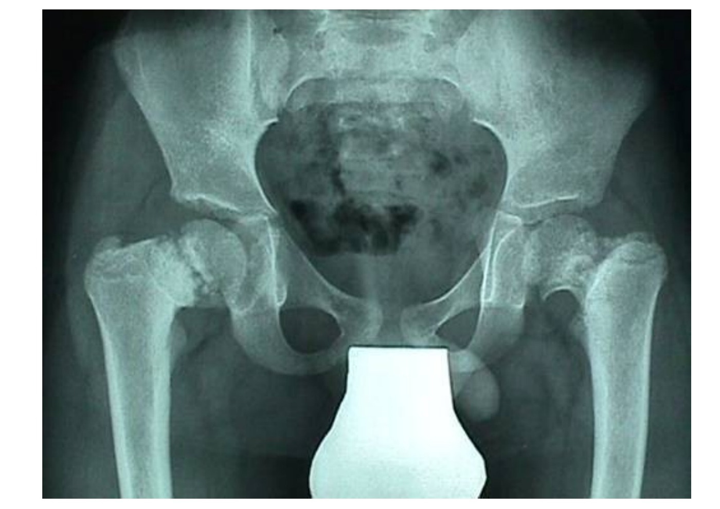
•Bilateral coxa vara deformity
•Vertical physis: Increased HE angle (Hilgenreiner epiphyseal angle) (>25 abnormal)
•Decreased neck shaft ankle (<125 abnormal)
•Coxa breva
•Inverted Y shape lucency in inf femoral neck (fairbank triangle) → ossification defect
•Acetabulum and tear drop normal
•Developmental coxa vara (AD)
Differential diagnosis
•Acquired: Perthes, chronic SCFE, trauma, infection
•Dysplasia: MED, SED, OI
•Metabolic: Cretinism (abnormal mental and physical development resulting from a deficiency of thyroid hormone in fetal or early life) Congenital: PFFD
•Bilateral in 30-50%
History
•Previous hip trauma or infection
•Associated skeletal abnormalities
Perinatal history
•FHx
Examination
•Trendelenburg gait
•LLD
•High riding GT
•Excessive lumbar lordosis
•Restrict hip ROM
Management
Prognosis by HE angle
•<45 20% resolve
•45-60 uncertain
•>60 will progress
Treatment is
•Observe
•VDRO (subtrochanteric <3 yo, intertroch >3yo) in HE > 60 , NSA <110
•CT preop – look for other associated deformities: coxa breva, femoral retroversion, acetabular dysplasia, GT overgrowth
•Goal: OVER correct NSA, correct LLD, correct version, restore abductor tension
Lateral approach
•Protect physis and periosteum
Aim HE <30
•Coxa vara = A decreased neck-shaft angle that is associated with an ossification defect in inferior femoral neck → vertical physis → increased shearing force