FRCS Paediatric Ortho — improved notes
FRCS Paediatric Ortho — visual inline notes

015. Perthes disease

visual-inline rebuild1 extracted figuresspeaker notes XML148 study lines16 Q&A

Individual slide images have been cropped from the rendered slide and inserted beside the matching note section. The full slide remains below only for layout/audit.

Clean FRCS notes with inline figures

Perthes disease

•Xray pelvis of skeletally immature patient with left hip pathology
•Left femoral proximal epiphysis appeared flattened and irregular, fragmented with lateral extrusion, cystic changes over metaphyseal area. The medial joint space is also widened. There is also lucency over lateral epiphysis, consistent with Gage sign.
•Contralateral hip not involved (10% bilateral, never same time)
•Working diagnosis would be Perthes disease. According to Waldenstrom classification, it looks to me the patient is in the fragmentation phase

*Waldenstrom classification

•Initial
•Fragmentation
•Reossification
•Remodeling
•Ddx are 1. septic arthritis, 2 Meyer’s disease 3. MED
•(bilateral Ddx: hypothyroid, lipid storage disease, meyers dysplasia, MED, sickle cell)
•Hx and PE would focus on
•Age, gender, body built
•Onset of symptoms
•Clinical at risk signs (FOOBS)
•R/O Ddx
•ROM (reduced abduction and IR, Trendelenburg gait, Catterall sign: hip flexion with obligatory external rotation)

Further investigations
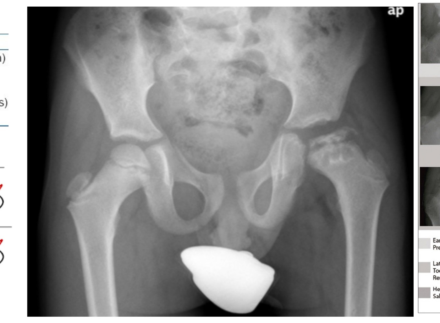
Radiograph / imaging - Perthes radiograph
Fig 1. Radiograph / imaging - Perthes radiographtop left
•WBC (infection), lipid (storage disorder), ESR, CRP, bone profile (tumour), TSH (hypothyroid), peripheral smear (sickle cell)

XR: Frog leg lateral

•+/- USG, aspiration if suspecting infection

Aim of treatment

•Pain control (rest analgesics)
•Maintain ROM (physio, temporary NWB)
•Contain hip during critical period of “biologic plasticity”, during creeping substitution, so the femoral head remodels with less incongruity (limits deformity and loss of sphericity) *abduction and IR*

Natural history:

•Classic Iowa study with a 50yr FU that 40% need arthroplasty if left untreated. Counsel patient regarding containment. Still has a possibility of future OA
•Nonsurgical containment: in abduction and IR

Indication for surgical containment

For this case:

Lateral pillar classification Class B or B/C

•>8yo treat surgically for containment→ improve outcome according to Herring’s study
•Proximal femoral varus osteotomy
•Prerequisites include
•- Timing – fragmentation phase
•- Containable
•→ Sx – good range
•→ XR – spherical and congruent reduction, containable upon abduction XR / intraop arthrogram

Operative steps

•Good preop plan to estimate the amount of varus needed to obtain good containment
•GA
•Adductor release may be indicated if tight

Lateral subvastus approach

•Periosteum elevated
•Choose appropriate size of paediatric proximal locking plate (3.5mm if <35kg, 5mm if >35kg)
•Set guiding jig based on plate angle and desired amount of varus (jig angle (current neck shaft angle) = plate angle (smaller)+ desired amount of varus produced)
•Fix plate proximally then remove it
•Mark rotation before osteotomy
•Use 2 k wires, one above and one below the osteotomy for rotational correction
•Perform femoral osteotomy using an oscillating saw
•Fix plate proximally then distally
•Add some internal rotation or extension for better containment
•Postop
•NWB walking
•HKAFO
•FU till maturity

Outcome depends on Stulberg classification:

•I normal
•II spherical congruent
•III non spherical (mushroom), congruent
•IV non spherical (concave articular surface) congruent
•V non spherical (flat), incongruent
•Other problems: LLD, trendelenburg, pain (labral tear, loose body, hinged abduction, FAI)

the etiology:

•1. Anatomical
•2. Hydrostatic pressure
•3. Thrombophilic

What is the blood supply to the femoral head in 4-9yo patients?

•<4yo: equal contribution by MFCA and LFCA
•4-10yo: LFCA regress and supply metaphysis only
•10-14yo: ligamentum teres contribution further decrease
•>14yo: anastomosis between the three systems (epiphyseal, metaphyseal, ligamentum teres)
•*What are the at risk sign (FOOBS)
•Clinical and radiological
•Clinical
•Female
•Old
•Obese
•Bilateral
•Stiffness

Radiological (Caterall at risk signs)

•Gage sign (inverted v shape radiolucency lateral to epiphysis)
•Horizontal physis
•Lateral subluxation of femoral head
•Calcification lateral to epiphysis
•Metaphyseal cyst (later added)

Lateral Pillar Classification (based on AP Xray, compare both sides, by Herring, guide management and prognosis)

•Lateral pillar: 25%, lateral to lucent line
•Central pillar: 50%
•Medial pillar: 20-35%
•A = normal height
•B = >50%
•B/C = exactly 50% + depressed relative to central pillar, very narrow lateral pillar >50% of original height, very little ossification but > 50% original height
•C = <50%

Treatment

Nonoperative containment

•Observation, activity restriction, NWB and ROM exercise (abd +IR)
•Petri cast. A frame (need to wear if for 6 months to 1 year) (problem, cannot guarantee abduction)

Operative containment

•1. Proximal femoral VDRO (varus) (at most 20deg) or ROWO (rot open wedge osteot.). Problems: short limb and Trendelenburg gait
•2. Pelvic osteotomy (for old child >8, as proximal femur does not remodel as much)
•Containable: re-directional (Salter) [problem: potential lengthening leading to increase pressure on head leading to AVN]
•Combination with femoral osteotomy increase coverage by 20%
•Shelf to prevent lateral subluxation / no longer containable,
•Chiari
•Valgus osteotomy for hinged abduction (reposition hinge, correct shortening from fixed adduction, improve abductor function)
•*** Advantage of surgical containment is the patient will go through fragmentation phase faster according to B Joseph

Herring’s study

•Prospective multi center study of 450 affected hips with patients of age 6+
•2004 JBJS

Prognosis depends on age and Herring stage

•A and B <8 did well regardless of treatment
•B, B/C > 8 improve outcome with surgery
•B/C <8, C poor regardless of treatment

no difference in outcomes between nonop treatment

•no difference in outcomes between femoral or acetabular osteotomies
•no significant effect regardless of treatment in those <8yo
•Limitation: AP view, 7 month of symptoms before reliable classification, 1/3 need upgrade grading, with only 4% belonging to group A on final grading, difficult in bilateral

*Benjamin Joseph’s study

•Containment aims at preventing or correcting femoral head extrusion
•Factors: age at onset of symptom, femoral head extrusion (>20% Reimer migration index), ROM
•B Joseph suggested containment ASAP for
•>7, contain ASAP if still in fragmentation phase
•If reduced ROM – restore motion by traction / abduction cast for 6 weeks, treat by containment once motion is restored
•<7, monitor and contain when there is presence of extrusion
•<8: Femoral osteotomy as more remodelling and less likely to have Trendelenburg gait later
•>8: Acetabular osteotomy as there will be less shortening.

Indication for containment surgery by BJ:

•>7 or <7 with extrusion
•Stage 1a 1b 2a (still in fragmentation phase)
•Extrusion present
•Normal ROM

Contraindication

•<7 without extrusion
•Stage 2b or above
•Absent extrusion in <7 years
•Restricted ROM

Healed Perthes:

Stulberg classification

•A la carte approach
•Trendelenburg: GT osteotomy
•Hinged abduction: osteochondroplasty
•Impingement, stiffness: Valgus osteotomy
•LLD
•Coxa brevia
•Coxa vara
•Coxa plana: Pelvis osteotomy

Direct Q&A

Q1 How is Perthes disease classified here?
•Initial
•Fragmentation
•Reossification
•Remodeling
•Ddx are 1. septic arthritis, 2 Meyer’s disease 3. MED
•(bilateral Ddx: hypothyroid, lipid storage disease, meyers dysplasia, MED, sickle cell)
•Hx and PE would focus on
•Age, gender, body built
•Onset of symptoms
•Clinical at risk signs (FOOBS)
•R/O Ddx
•ROM (reduced abduction and IR, Trendelenburg gait, Catterall sign: hip flexion with obligatory external rotation)
Q2 What investigations are listed for Perthes disease?
•WBC (infection), lipid (storage disorder), ESR, CRP, bone profile (tumour), TSH (hypothyroid), peripheral smear (sickle cell)
Q3 What are the radiographic findings for Perthes disease?

+/- USG, aspiration if suspecting infection

Q4 What are the management aims for Perthes disease?

Pain control (rest analgesics)

•Maintain ROM (physio, temporary NWB)
•Contain hip during critical period of “biologic plasticity”, during creeping substitution, so the femoral head remodels with less incongruity (limits deformity and loss of sphericity) *abduction and IR*
Q5 What natural history is described for Perthes disease?
•Classic Iowa study with a 50yr FU that 40% need arthroplasty if left untreated. Counsel patient regarding containment. Still has a possibility of future OA

Nonsurgical containment: in abduction and IR

Q6 What operative steps, indications, and risks are listed for Operative steps?
•Good preop plan to estimate the amount of varus needed to obtain good containment
•GA
•Adductor release may be indicated if tight
Q7 What operative steps, indications, and risks are listed for Lateral subvastus approach?

Periosteum elevated

•Choose appropriate size of paediatric proximal locking plate (3.5mm if <35kg, 5mm if >35kg)
•Set guiding jig based on plate angle and desired amount of varus (jig angle (current neck shaft angle) = plate angle (smaller)+ desired amount of varus produced)
•Fix plate proximally then remove it
•Mark rotation before osteotomy
•Use 2 k wires, one above and one below the osteotomy for rotational correction
•Perform femoral osteotomy using an oscillating saw
•Fix plate proximally then distally
•Add some internal rotation or extension for better containment
•Postop
•NWB walking
•HKAFO
•FU till maturity
Q8 What aetiology is described for Perthes disease?
•1. Anatomical
•2. Hydrostatic pressure
•3. Thrombophilic
Q9 What is the blood supply to the femoral head in 4-9yo patients?
•<4yo: equal contribution by MFCA and LFCA
•4-10yo: LFCA regress and supply metaphysis only
•10-14yo: ligamentum teres contribution further decrease
•>14yo: anastomosis between the three systems (epiphyseal, metaphyseal, ligamentum teres)
•*What are the at risk sign (FOOBS)
•Clinical and radiological
•Clinical
•Female
•Old
•Obese
•Bilateral
•Stiffness
Q10 What signs or features are listed for Perthes disease?

Gage sign (inverted v shape radiolucency lateral to epiphysis)

•Horizontal physis
•Lateral subluxation of femoral head
•Calcification lateral to epiphysis
•Metaphyseal cyst (later added)
Q11 What operative steps, indications, and risks are listed for Nonoperative containment?

Observation, activity restriction, NWB and ROM exercise (abd +IR)

•Petri cast. A frame (need to wear if for 6 months to 1 year) (problem, cannot guarantee abduction)
Q12 What operative steps, indications, and risks are listed for Operative containment?
•1. Proximal femoral VDRO (varus) (at most 20deg) or ROWO (rot open wedge osteot.). Problems: short limb and Trendelenburg gait
•2. Pelvic osteotomy (for old child >8, as proximal femur does not remodel as much)
•Containable: re-directional (Salter) [problem: potential lengthening leading to increase pressure on head leading to AVN]
•Combination with femoral osteotomy increase coverage by 20%
•Shelf to prevent lateral subluxation / no longer containable,
•Chiari
•Valgus osteotomy for hinged abduction (reposition hinge, correct shortening from fixed adduction, improve abductor function)
•*** Advantage of surgical containment is the patient will go through fragmentation phase faster according to B Joseph
Q13 What evidence or study finding is cited for Perthes disease?
•Prospective multi center study of 450 affected hips with patients of age 6+
•2004 JBJS
Q14 What thresholds, cut-offs, or criteria are listed under no difference in outcomes between nonop treatment?

no difference in outcomes between femoral or acetabular osteotomies

•no significant effect regardless of treatment in those <8yo
•Limitation: AP view, 7 month of symptoms before reliable classification, 1/3 need upgrade grading, with only 4% belonging to group A on final grading, difficult in bilateral
Q15 What operative steps, indications, and risks are listed for Indication for containment surgery by BJ?
•>7 or <7 with extrusion
•Stage 1a 1b 2a (still in fragmentation phase)
•Extrusion present
•Normal ROM
Q16 What are the indications listed for Perthes disease?
•<7 without extrusion
•Stage 2b or above
•Absent extrusion in <7 years
•Restricted ROM

Raw speaker-note hierarchy audit

#lvlmarLindentlogicaloriginal note line
10NoneNone0Perthes disease
20NoneNone0Xray pelvis of skeletally immature patient with left hip pathology
30NoneNone0Left femoral proximal epiphysis appeared flattened and irregular, fragmented with lateral extrusion, cystic changes over metaphyseal area. The medial joint space is also widened. There is also lucency over lateral epiphysis, consistent with Gage sign.
40NoneNone0Contralateral hip not involved (10% bilateral, never same time)
50NoneNone0Working diagnosis would be Perthes disease. According to Waldenstrom classification, it looks to me the patient is in the fragmentation phase
60NoneNone0*Waldenstrom classification
71685800-2286002Initial
81685800-2286002Fragmentation
91685800-2286002Reossification
101685800-2286002Remodeling
110NoneNone0Ddx are 1. septic arthritis, 2 Meyer’s disease 3. MED
120NoneNone0(bilateral Ddx: hypothyroid, lipid storage disease, meyers dysplasia, MED, sickle cell)
130NoneNone0Hx and PE would focus on
140228600-2286001Age, gender, body built
150228600-2286001Onset of symptoms
160228600-2286001Clinical at risk signs (FOOBS)
170228600-2286001R/O Ddx
180228600-2286001ROM (reduced abduction and IR, Trendelenburg gait, Catterall sign: hip flexion with obligatory external rotation)
190NoneNone0Further Investigation
200NoneNone0WBC (infection), lipid (storage disorder), ESR, CRP, bone profile (tumour), TSH (hypothyroid), peripheral smear (sickle cell)
210NoneNone0XR: Frog leg lateral
220NoneNone0+/- USG, aspiration if suspecting infection
230NoneNone0Aim of treatment
240228600-2286001Pain control (rest analgesics)
250228600-2286001Maintain ROM (physio, temporary NWB)
260228600-2286001Contain hip during critical period of “biologic plasticity”, during creeping substitution, so the femoral head remodels with less incongruity (limits deformity and loss of sphericity) *abduction and IR*
270000Natural history:
280000Classic Iowa study with a 50yr FU that 40% need arthroplasty if left untreated. Counsel patient regarding containment. Still has a possibility of future OA
290000Nonsurgical containment: in abduction and IR
300000Indication for surgical containment
310000For this case:
320NoneNone0Lateral pillar classification Class B or B/C
330NoneNone0>8yo treat surgically for containment→ improve outcome according to Herring’s study
340NoneNone0Proximal femoral varus osteotomy
350NoneNone0Prerequisites include
360000- Timing – fragmentation phase
370000- Containable
380000→ Sx – good range
390NoneNone0→ XR – spherical and congruent reduction, containable upon abduction XR / intraop arthrogram
400NoneNone0Take me through the surgery
410228600-2286001Good preop plan to estimate the amount of varus needed to obtain good containment
420228600-2286001GA
430228600-2286001Adductor release may be indicated if tight
440228600-2286001Lateral subvastus approach
450228600-2286001Periosteum elevated
460228600-2286001Choose appropriate size of paediatric proximal locking plate (3.5mm if <35kg, 5mm if >35kg)
470228600-2286001Set guiding jig based on plate angle and desired amount of varus (jig angle (current neck shaft angle) = plate angle (smaller)+ desired amount of varus produced)
480228600-2286001Fix plate proximally then remove it
490228600-2286001Mark rotation before osteotomy
500228600-2286001Use 2 k wires, one above and one below the osteotomy for rotational correction
510228600-2286001Perform femoral osteotomy using an oscillating saw
520228600-2286001Fix plate proximally then distally
530228600-2286001Add some internal rotation or extension for better containment
540NoneNone0Postop
550NoneNone0NWB walking
560NoneNone0HKAFO
570NoneNone0FU till maturity
580000Outcome depends on Stulberg classification:
59145720001I normal
60145720001II spherical congruent
61145720001III non spherical (mushroom), congruent
62145720001IV non spherical (concave articular surface) congruent
63145720001V non spherical (flat), incongruent
640NoneNone0Other problems: LLD, trendelenburg, pain (labral tear, loose body, hinged abduction, FAI)
650NoneNone0What is the etiology:
660NoneNone01. Anatomical
670NoneNone02. Hydrostatic pressure
680NoneNone03. Thrombophilic
690NoneNone0What is the blood supply to the femoral head in 4-9yo patients?
701NoneNone1<4yo: equal contribution by MFCA and LFCA
711NoneNone14-10yo: LFCA regress and supply metaphysis only
721NoneNone110-14yo: ligamentum teres contribution further decrease
731NoneNone1>14yo: anastomosis between the three systems (epiphyseal, metaphyseal, ligamentum teres)
740NoneNone0*What are the at risk sign (FOOBS)
750NoneNone0Clinical and radiological
760NoneNone0Clinical
771685800-2286002Female
781685800-2286002Old
791685800-2286002Obese
801685800-2286002Bilateral
811685800-2286002Stiffness
820NoneNone0Radiological (Caterall at risk signs)
831685800-2286002Gage sign (inverted v shape radiolucency lateral to epiphysis)
841685800-2286002Horizontal physis
851685800-2286002Lateral subluxation of femoral head
861685800-2286002Calcification lateral to epiphysis
871685800-2286002Metaphyseal cyst (later added)
880000Lateral Pillar Classification (based on AP Xray, compare both sides, by Herring, guide management and prognosis)
89145720001Lateral pillar: 25%, lateral to lucent line
90145720001Central pillar: 50%
91145720001Medial pillar: 20-35%
920171450-1714501A = normal height
930171450-1714501B = >50%
940171450-1714501B/C = exactly 50% + depressed relative to central pillar, very narrow lateral pillar >50% of original height, very little ossification but > 50% original height
950171450-1714501C = <50%
960000Treatment
970000Nonoperative containment
980000Observation, activity restriction, NWB and ROM exercise (abd +IR)
990000Petri cast. A frame (need to wear if for 6 months to 1 year) (problem, cannot guarantee abduction)
1000000Operative containment
10100001. Proximal femoral VDRO (varus) (at most 20deg) or ROWO (rot open wedge osteot.). Problems: short limb and Trendelenburg gait
10200002. Pelvic osteotomy (for old child >8, as proximal femur does not remodel as much)
1030171450-1714501Containable: re-directional (Salter) [problem: potential lengthening leading to increase pressure on head leading to AVN]
1040171450-1714501Combination with femoral osteotomy increase coverage by 20%
1050171450-1714501Shelf to prevent lateral subluxation / no longer containable,
1060171450-1714501Chiari
1070171450-1714501Valgus osteotomy for hinged abduction (reposition hinge, correct shortening from fixed adduction, improve abductor function)
1080000*** Advantage of surgical containment is the patient will go through fragmentation phase faster according to B Joseph
1090000Tell me about Herring’s study
1100000Prospective multi center study of 450 affected hips with patients of age 6+
11100002004 JBJS
1120000Prognosis depends on age and Herring stage
113145720001A and B <8 did well regardless of treatment
114145720001B, B/C > 8 improve outcome with surgery
115145720001B/C <8, C poor regardless of treatment
1160171450-1714501no difference in outcomes between nonop treatment
1170171450-1714501no difference in outcomes between femoral or acetabular osteotomies
1180171450-1714501no significant effect regardless of treatment in those <8yo
1190000Limitation: AP view, 7 month of symptoms before reliable classification, 1/3 need upgrade grading, with only 4% belonging to group A on final grading, difficult in bilateral
1200000*Benjamin Joseph’s study
1210000Containment aims at preventing or correcting femoral head extrusion
1220000Factors: age at onset of symptom, femoral head extrusion (>20% Reimer migration index), ROM
1230000B Joseph suggested containment ASAP for
1240171450-1714501>7, contain ASAP if still in fragmentation phase
1251628650-1714502If reduced ROM – restore motion by traction / abduction cast for 6 weeks, treat by containment once motion is restored
1260171450-1714501<7, monitor and contain when there is presence of extrusion
1270171450-1714501<8: Femoral osteotomy as more remodelling and less likely to have Trendelenburg gait later
1280171450-1714501>8: Acetabular osteotomy as there will be less shortening.
1290000Indication for containment surgery by BJ:
1300228600-2286001>7 or <7 with extrusion
1310228600-2286001Stage 1a 1b 2a (still in fragmentation phase)
1320228600-2286001Extrusion present
1330228600-2286001Normal ROM
1340000Contraindication
1350228600-2286001<7 without extrusion
1360228600-2286001Stage 2b or above
1370228600-2286001Absent extrusion in <7 years
1380228600-2286001Restricted ROM
1390000Healed Perthes:
1400000Stulberg classification
1410000A la carte approach
1421628650-1714502Trendelenburg: GT osteotomy
1431628650-1714502Hinged abduction: osteochondroplasty
1441628650-1714502Impingement, stiffness: Valgus osteotomy
1451628650-1714502LLD
1461628650-1714502Coxa brevia
1471628650-1714502Coxa vara
1481628650-1714502Coxa plana: Pelvis osteotomy

Improved semantic hierarchy used for the study notes

#semantic levelsection?rewritten line
10Perthes disease
21Xray pelvis of skeletally immature patient with left hip pathology
31Left femoral proximal epiphysis appeared flattened and irregular, fragmented with lateral extrusion, cystic changes over metaphyseal area. The medial joint space is also widened. There is also lucency over lateral epiphysis, consistent with Gage sign.
41Contralateral hip not involved (10% bilateral, never same time)
51Working diagnosis would be Perthes disease. According to Waldenstrom classification, it looks to me the patient is in the fragmentation phase
60yes*Waldenstrom classification
72Initial
82Fragmentation
92Reossification
102Remodeling
111Ddx are 1. septic arthritis, 2 Meyer’s disease 3. MED
121(bilateral Ddx: hypothyroid, lipid storage disease, meyers dysplasia, MED, sickle cell)
131Hx and PE would focus on
141Age, gender, body built
151Onset of symptoms
161Clinical at risk signs (FOOBS)
171R/O Ddx
181ROM (reduced abduction and IR, Trendelenburg gait, Catterall sign: hip flexion with obligatory external rotation)
190yesFurther investigations
201WBC (infection), lipid (storage disorder), ESR, CRP, bone profile (tumour), TSH (hypothyroid), peripheral smear (sickle cell)
210yesXR: Frog leg lateral
221+/- USG, aspiration if suspecting infection
230yesAim of treatment
241Pain control (rest analgesics)
251Maintain ROM (physio, temporary NWB)
261Contain hip during critical period of “biologic plasticity”, during creeping substitution, so the femoral head remodels with less incongruity (limits deformity and loss of sphericity) *abduction and IR*
270yesNatural history:
281Classic Iowa study with a 50yr FU that 40% need arthroplasty if left untreated. Counsel patient regarding containment. Still has a possibility of future OA
291Nonsurgical containment: in abduction and IR
300yesIndication for surgical containment
310yesFor this case:
320yesLateral pillar classification Class B or B/C
331>8yo treat surgically for containment→ improve outcome according to Herring’s study
341Proximal femoral varus osteotomy
351Prerequisites include
361- Timing – fragmentation phase
371- Containable
381→ Sx – good range
391→ XR – spherical and congruent reduction, containable upon abduction XR / intraop arthrogram
400yesOperative steps
411Good preop plan to estimate the amount of varus needed to obtain good containment
421GA
431Adductor release may be indicated if tight
440yesLateral subvastus approach
451Periosteum elevated
461Choose appropriate size of paediatric proximal locking plate (3.5mm if <35kg, 5mm if >35kg)
471Set guiding jig based on plate angle and desired amount of varus (jig angle (current neck shaft angle) = plate angle (smaller)+ desired amount of varus produced)
481Fix plate proximally then remove it
491Mark rotation before osteotomy
501Use 2 k wires, one above and one below the osteotomy for rotational correction
511Perform femoral osteotomy using an oscillating saw
521Fix plate proximally then distally
531Add some internal rotation or extension for better containment
541Postop
551NWB walking
561HKAFO
571FU till maturity
580yesOutcome depends on Stulberg classification:
591I normal
601II spherical congruent
611III non spherical (mushroom), congruent
621IV non spherical (concave articular surface) congruent
631V non spherical (flat), incongruent
641Other problems: LLD, trendelenburg, pain (labral tear, loose body, hinged abduction, FAI)
650yesthe etiology:
6611. Anatomical
6712. Hydrostatic pressure
6813. Thrombophilic
690yesWhat is the blood supply to the femoral head in 4-9yo patients?
701<4yo: equal contribution by MFCA and LFCA
7114-10yo: LFCA regress and supply metaphysis only
72110-14yo: ligamentum teres contribution further decrease
731>14yo: anastomosis between the three systems (epiphyseal, metaphyseal, ligamentum teres)
741*What are the at risk sign (FOOBS)
751Clinical and radiological
761Clinical
772Female
782Old
792Obese
802Bilateral
812Stiffness
820yesRadiological (Caterall at risk signs)
832Gage sign (inverted v shape radiolucency lateral to epiphysis)
842Horizontal physis
852Lateral subluxation of femoral head
862Calcification lateral to epiphysis
872Metaphyseal cyst (later added)
880yesLateral Pillar Classification (based on AP Xray, compare both sides, by Herring, guide management and prognosis)
891Lateral pillar: 25%, lateral to lucent line
901Central pillar: 50%
911Medial pillar: 20-35%
921A = normal height
931B = >50%
941B/C = exactly 50% + depressed relative to central pillar, very narrow lateral pillar >50% of original height, very little ossification but > 50% original height
951C = <50%
960yesTreatment
970yesNonoperative containment
981Observation, activity restriction, NWB and ROM exercise (abd +IR)
991Petri cast. A frame (need to wear if for 6 months to 1 year) (problem, cannot guarantee abduction)
1000yesOperative containment
10111. Proximal femoral VDRO (varus) (at most 20deg) or ROWO (rot open wedge osteot.). Problems: short limb and Trendelenburg gait
10212. Pelvic osteotomy (for old child >8, as proximal femur does not remodel as much)
1031Containable: re-directional (Salter) [problem: potential lengthening leading to increase pressure on head leading to AVN]
1041Combination with femoral osteotomy increase coverage by 20%
1051Shelf to prevent lateral subluxation / no longer containable,
1061Chiari
1071Valgus osteotomy for hinged abduction (reposition hinge, correct shortening from fixed adduction, improve abductor function)
1081*** Advantage of surgical containment is the patient will go through fragmentation phase faster according to B Joseph
1090yesHerring’s study
1101Prospective multi center study of 450 affected hips with patients of age 6+
11112004 JBJS
1120yesPrognosis depends on age and Herring stage
1131A and B <8 did well regardless of treatment
1141B, B/C > 8 improve outcome with surgery
1151B/C <8, C poor regardless of treatment
1160yesno difference in outcomes between nonop treatment
1171no difference in outcomes between femoral or acetabular osteotomies
1181no significant effect regardless of treatment in those <8yo
1191Limitation: AP view, 7 month of symptoms before reliable classification, 1/3 need upgrade grading, with only 4% belonging to group A on final grading, difficult in bilateral
1200yes*Benjamin Joseph’s study
1211Containment aims at preventing or correcting femoral head extrusion
1221Factors: age at onset of symptom, femoral head extrusion (>20% Reimer migration index), ROM
1231B Joseph suggested containment ASAP for
1241>7, contain ASAP if still in fragmentation phase
1252If reduced ROM – restore motion by traction / abduction cast for 6 weeks, treat by containment once motion is restored
1261<7, monitor and contain when there is presence of extrusion
1271<8: Femoral osteotomy as more remodelling and less likely to have Trendelenburg gait later
1281>8: Acetabular osteotomy as there will be less shortening.
1290yesIndication for containment surgery by BJ:
1301>7 or <7 with extrusion
1311Stage 1a 1b 2a (still in fragmentation phase)
1321Extrusion present
1331Normal ROM
1340yesContraindication
1351<7 without extrusion
1361Stage 2b or above
1371Absent extrusion in <7 years
1381Restricted ROM
1390yesHealed Perthes:
1400yesStulberg classification
1411A la carte approach
1422Trendelenburg: GT osteotomy
1432Hinged abduction: osteochondroplasty
1442Impingement, stiffness: Valgus osteotomy
1452LLD
1462Coxa brevia
1472Coxa vara
1482Coxa plana: Pelvis osteotomy