FRCS Paediatric Ortho — improved notes
FRCS Paediatric Ortho — visual inline notes

019. Congenital radioulnar synostosis

visual-inline rebuild2 extracted figuresspeaker notes XML71 study lines7 Q&A

Individual slide images have been cropped from the rendered slide and inserted beside the matching note section. The full slide remains below only for layout/audit.

Clean FRCS notes with inline figures

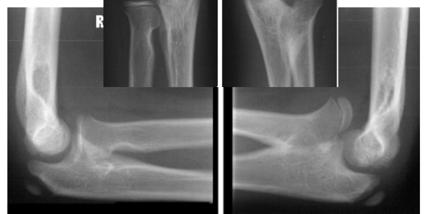
Xray elbow of skeletally immature patient
Radiograph / imaging - radioulnar synostosis radiograph
Fig 1. Radiograph / imaging - radioulnar synostosis radiographmiddle left
Radiograph / imaging - radioulnar synostosis radiograph
Fig 2. Radiograph / imaging - radioulnar synostosis radiographtop left
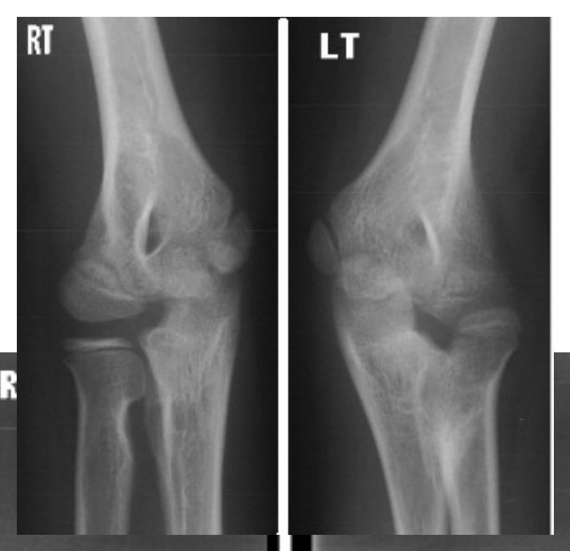
•Radioulnar synostosis bilaterally
•Left ant dislocated radial head with malformation
•Likely congenital as it is bilateral and there is continuity of IM canal and matrix
•Ddx: acquire RU synostosis, radiohumeral synostosis
•(AD with variable penetrance, Failure of segmentation, 60% bilateral)
•Cleary classification type I on right, type IV on left
•[may also be given a clinical photo: need to comment on asymmetry in resting position, any compensation with shoulder adduction and also presence of elbow crease]

History

History of forearm fracture

•FHx
•Current disability
•Bilateral

Examination

•See if there is any forearm bowing
•Assess function (depends on fused position)
•Most are in pronation (>50% >50deg pronation)

Look for other associations:

•Apert syndrome (acrocephlosyndactyly)
•Carpenter’s syndrome (acropolysyndactyly)
•Arthrogryposis
•Klinefelter’s syndrome
•Xray whole forearm and chromosome analysis (some have duplication in sex-chromosome – Klinefelter syndrome)

Management

•No intervention if ADL coped well (usu. wrist more mobile than normal)

Indications for surgery:

•Functional deficit (pronation >60 degree)

Time of surgery – 5yo

•Rotational osteotomy at level of synostosis
•Large contact area, less compartment syndrome, along axis of rotation, high power of correction
•- Prophylactic fasciotomy and PIN nerve release
•At most 80deg correction in each OT

Position depends on demand

•Unilateral: 20’ pronation (or neutral)
•Bilateral: Dominant hand : 30-45‘ pronation, Non-dominant hand : 20-35’ supination
•Cx: PIN injury, compartment syndrome
•(Resection have high chance of recurrence)

Cleary classification

Type / groupDefinition / note
1No osseous synostosis, radial head ok
2Osseous synostosis, radial head ok
3Long osseous synostosis, radial head posterior dislocated
4short osseous synostosis, radial head anterior dislocated
•Pathogenesis: failed segmentation
•Longitudinal segmentation starts at 6 weeks from distal to proximal
•Forearm at pronation at the time (limb rotation complete at 9 weeks)
•Always at pronation → radioulnar growth not in proportion → radial head dislocation
•C/f Acquired (post traumatic)
•Congenital always proximal due to division from distal to proximal
•Continuity of IM canal
•60% bilateral
•Radial head dislocation: mushroom radial head, hypoplastic capitellum, long radial neck, radial bow rather than ulnar bow (old monteggia)

Risk factor of posttraumatic synostosis :

•Injury factor: 1 # both bones at same level, 2 crush/ burn, 3 head injury
•Intervention factor: 1 surgical delay >2 weeks 2 single incision 3 disruption of IO membrane 4 infection
•→ Rx: physio, RT, NSAID, proximal radial excision
•the embryological development of limb
•4 weeks: Limb bud appearance from 8th to 10th somites as an outpouching composing of mesoderm covered by ectoderm. Formation of anlage.
•5 weeks: Formation of neural plexus
•6 weeks:
•Differentiation of mesenchyme into cartilage (chondrification)
•Segmentation (formation of primitive joint)
•7 weeks: formation of primary ossification centre (complete by 12 weeks)
•Intramembranous ossification at periosteal sleeve
•Enchondral ossification at central hypertrophied cartilage
•9 weeks: limbs rotate (UL90 ER, LL 90 IR)
•36 weeks: formation of secondary ossification centre
•3 axis of limb patterning
•Apical epidermal ridge (AER) – proximodistal (HOX gene, secrete Fbl growth factor) (e.g. radial club hand)
•Zone of polarizing activity – AP (secrete Shh protein) [ Mirror hand ]
•Progress zone: Wnt pathway: Dorsoventral (secret LMX1) [e.g. nail patella]

Direct Q&A

Q1 What are the radiographic findings for Congenital radioulnar synostosis?

Radioulnar synostosis bilaterally

•Left ant dislocated radial head with malformation
•Likely congenital as it is bilateral and there is continuity of IM canal and matrix
•Ddx: acquire RU synostosis, radiohumeral synostosis
•(AD with variable penetrance, Failure of segmentation, 60% bilateral)

Cleary classification type I on right, type IV on left

•[may also be given a clinical photo: need to comment on asymmetry in resting position, any compensation with shoulder adduction and also presence of elbow crease]
Q2 What examination findings should be assessed in Congenital radioulnar synostosis?

See if there is any forearm bowing

•Assess function (depends on fused position)
•Most are in pronation (>50% >50deg pronation)
Q3 How is Congenital radioulnar synostosis managed?
•No intervention if ADL coped well (usu. wrist more mobile than normal)
Q4 What operative steps, indications, and risks are listed for Indications for surgery?
•Functional deficit (pronation >60 degree)
Q5 What operative steps, indications, and risks are listed for Time of surgery – 5yo?

Rotational osteotomy at level of synostosis

•Large contact area, less compartment syndrome, along axis of rotation, high power of correction
•- Prophylactic fasciotomy and PIN nerve release
•At most 80deg correction in each OT
Q6 How is Congenital radioulnar synostosis classified here?

1: No osseous synostosis, radial head ok

•2: Osseous synostosis, radial head ok
•3: Long osseous synostosis, radial head posterior dislocated
•4: short osseous synostosis, radial head anterior dislocated

Pathogenesis: failed segmentation

•Longitudinal segmentation starts at 6 weeks from distal to proximal
•Forearm at pronation at the time (limb rotation complete at 9 weeks)
•Always at pronation → radioulnar growth not in proportion → radial head dislocation
•C/f Acquired (post traumatic)
•Congenital always proximal due to division from distal to proximal
•Continuity of IM canal
•60% bilateral
•Radial head dislocation: mushroom radial head, hypoplastic capitellum, long radial neck, radial bow rather than ulnar bow (old monteggia)
Q7 What risk factors are listed for Congenital radioulnar synostosis?
•Injury factor: 1 # both bones at same level, 2 crush/ burn, 3 head injury
•Intervention factor: 1 surgical delay >2 weeks 2 single incision 3 disruption of IO membrane 4 infection
•→ Rx: physio, RT, NSAID, proximal radial excision
•the embryological development of limb
•4 weeks: Limb bud appearance from 8th to 10th somites as an outpouching composing of mesoderm covered by ectoderm. Formation of anlage.
•5 weeks: Formation of neural plexus
•6 weeks:
•Differentiation of mesenchyme into cartilage (chondrification)
•Segmentation (formation of primitive joint)
•7 weeks: formation of primary ossification centre (complete by 12 weeks)
•Intramembranous ossification at periosteal sleeve
•Enchondral ossification at central hypertrophied cartilage
•9 weeks: limbs rotate (UL90 ER, LL 90 IR)
•36 weeks: formation of secondary ossification centre
•3 axis of limb patterning
•Apical epidermal ridge (AER) – proximodistal (HOX gene, secrete Fbl growth factor) (e.g. radial club hand)
•Zone of polarizing activity – AP (secrete Shh protein) [ Mirror hand ]
•Progress zone: Wnt pathway: Dorsoventral (secret LMX1) [e.g. nail patella]

Raw speaker-note hierarchy audit

#lvlmarLindentlogicaloriginal note line
10NoneNone0Xray elbow of skeletally immature patient
20NoneNone0Radioulnar synostosis bilaterally
30NoneNone0Left ant dislocated radial head with malformation
40NoneNone0Likely congenital as it is bilateral and there is continuity of IM canal and matrix
50NoneNone0Ddx: acquire RU synostosis, radiohumeral synostosis
60NoneNone0(AD with variable penetrance, Failure of segmentation, 60% bilateral)
70NoneNone0Cleary classification type I on right, type IV on left
80NoneNone0[may also be given a clinical photo: need to comment on asymmetry in resting position, any compensation with shoulder adduction and also presence of elbow crease]
90NoneNone0Hx:
100NoneNone0History of forearm fracture
110NoneNone0FHx
120NoneNone0Current disability
130NoneNone0Bilateral
140NoneNone0PE
150NoneNone0See if there is any forearm bowing
160000Assess function (depends on fused position)
170NoneNone0Most are in pronation (>50% >50deg pronation)
180NoneNone0Look for other associations:
191628650-1714502Apert syndrome (acrocephlosyndactyly)
201628650-1714502Carpenter’s syndrome (acropolysyndactyly)
211628650-1714502Arthrogryposis
221628650-1714502Klinefelter’s syndrome
230NoneNone0Xray whole forearm and chromosome analysis (some have duplication in sex-chromosome – Klinefelter syndrome)
240NoneNone0Mx:
250NoneNone0No intervention if ADL coped well (usu. wrist more mobile than normal)
260NoneNone0Indications for surgery:
270171450-1714501Functional deficit (pronation >60 degree)
280000Time of surgery – 5yo
290NoneNone0Rotational osteotomy at level of synostosis
300NoneNone0Large contact area, less compartment syndrome, along axis of rotation, high power of correction
310NoneNone0- Prophylactic fasciotomy and PIN nerve release
320171450-1714501At most 80deg correction in each OT
330171450-1714501Position depends on demand
341628650-1714502Unilateral: 20’ pronation (or neutral)
351628650-1714502Bilateral: Dominant hand : 30-45‘ pronation, Non-dominant hand : 20-35’ supination
360NoneNone0Cx: PIN injury, compartment syndrome
370NoneNone0(Resection have high chance of recurrence)
380NoneNone0=======================================================
390NoneNone0Cleary classifcation
400NoneNone01: No osseous synostosis, radial head ok
410NoneNone02: Osseous synostosis, radial head ok
420NoneNone03: Long osseous synostosis, radial head posterior dislocated
430NoneNone04: short osseous synostosis, radial head anterior dislocated
440NoneNone0Pathogenesis: failed segmentation
450171450-1714501Longitudinal segmentation starts at 6 weeks from distal to proximal
460171450-1714501Forearm at pronation at the time (limb rotation complete at 9 weeks)
470171450-1714501Always at pronation → radioulnar growth not in proportion → radial head dislocation
480NoneNone0C/f Acquired (post traumatic)
490342900-3429001Congenital always proximal due to division from distal to proximal
500342900-3429001Continuity of IM canal
510342900-342900160% bilateral
520342900-3429001Radial head dislocation: mushroom radial head, hypoplastic capitellum, long radial neck, radial bow rather than ulnar bow (old monteggia)
530000Risk factor of posttraumatic synostosis :
540171450-1714501Injury factor: 1 # both bones at same level, 2 crush/ burn, 3 head injury
550171450-1714501Intervention factor: 1 surgical delay >2 weeks 2 single incision 3 disruption of IO membrane 4 infection
560000→ Rx: physio, RT, NSAID, proximal radial excision
570NoneNone0Tell me about the embryological development of limb
580171450-17145014 weeks: Limb bud appearance from 8th to 10th somites as an outpouching composing of mesoderm covered by ectoderm. Formation of anlage.
590171450-17145015 weeks: Formation of neural plexus
600171450-17145016 weeks:
611628650-1714502Differentiation of mesenchyme into cartilage (chondrification)
621628650-1714502Segmentation (formation of primitive joint)
630171450-17145017 weeks: formation of primary ossification centre (complete by 12 weeks)
641628650-1714502Intramembranous ossification at periosteal sleeve
651628650-1714502Enchondral ossification at central hypertrophied cartilage
660171450-17145019 weeks: limbs rotate (UL90 ER, LL 90 IR)
670171450-171450136 weeks: formation of secondary ossification centre
680NoneNone03 axis of limb patterning
691NoneNone1Apical epidermal ridge (AER) – proximodistal (HOX gene, secrete Fbl growth factor) (e.g. radial club hand)
701NoneNone1Zone of polarizing activity – AP (secrete Shh protein) [ Mirror hand ]
711NoneNone1Progress zone: Wnt pathway: Dorsoventral (secret LMX1) [e.g. nail patella]

Improved semantic hierarchy used for the study notes

#semantic levelsection?rewritten line
10yesXray elbow of skeletally immature patient
21Radioulnar synostosis bilaterally
31Left ant dislocated radial head with malformation
41Likely congenital as it is bilateral and there is continuity of IM canal and matrix
51Ddx: acquire RU synostosis, radiohumeral synostosis
61(AD with variable penetrance, Failure of segmentation, 60% bilateral)
71Cleary classification type I on right, type IV on left
81[may also be given a clinical photo: need to comment on asymmetry in resting position, any compensation with shoulder adduction and also presence of elbow crease]
90yesHistory
100yesHistory of forearm fracture
111FHx
121Current disability
131Bilateral
140yesExamination
151See if there is any forearm bowing
161Assess function (depends on fused position)
171Most are in pronation (>50% >50deg pronation)
180yesLook for other associations:
192Apert syndrome (acrocephlosyndactyly)
202Carpenter’s syndrome (acropolysyndactyly)
212Arthrogryposis
222Klinefelter’s syndrome
231Xray whole forearm and chromosome analysis (some have duplication in sex-chromosome – Klinefelter syndrome)
240yesManagement
251No intervention if ADL coped well (usu. wrist more mobile than normal)
260yesIndications for surgery:
271Functional deficit (pronation >60 degree)
280yesTime of surgery – 5yo
291Rotational osteotomy at level of synostosis
301Large contact area, less compartment syndrome, along axis of rotation, high power of correction
311- Prophylactic fasciotomy and PIN nerve release
321At most 80deg correction in each OT
330yesPosition depends on demand
342Unilateral: 20’ pronation (or neutral)
352Bilateral: Dominant hand : 30-45‘ pronation, Non-dominant hand : 20-35’ supination
361Cx: PIN injury, compartment syndrome
371(Resection have high chance of recurrence)
380——
390yesCleary classification
4011: No osseous synostosis, radial head ok
4112: Osseous synostosis, radial head ok
4213: Long osseous synostosis, radial head posterior dislocated
4314: short osseous synostosis, radial head anterior dislocated
441Pathogenesis: failed segmentation
451Longitudinal segmentation starts at 6 weeks from distal to proximal
461Forearm at pronation at the time (limb rotation complete at 9 weeks)
471Always at pronation → radioulnar growth not in proportion → radial head dislocation
481C/f Acquired (post traumatic)
491Congenital always proximal due to division from distal to proximal
501Continuity of IM canal
51160% bilateral
521Radial head dislocation: mushroom radial head, hypoplastic capitellum, long radial neck, radial bow rather than ulnar bow (old monteggia)
530yesRisk factor of posttraumatic synostosis :
541Injury factor: 1 # both bones at same level, 2 crush/ burn, 3 head injury
551Intervention factor: 1 surgical delay >2 weeks 2 single incision 3 disruption of IO membrane 4 infection
561→ Rx: physio, RT, NSAID, proximal radial excision
571the embryological development of limb
5814 weeks: Limb bud appearance from 8th to 10th somites as an outpouching composing of mesoderm covered by ectoderm. Formation of anlage.
5915 weeks: Formation of neural plexus
6016 weeks:
612Differentiation of mesenchyme into cartilage (chondrification)
622Segmentation (formation of primitive joint)
6317 weeks: formation of primary ossification centre (complete by 12 weeks)
642Intramembranous ossification at periosteal sleeve
652Enchondral ossification at central hypertrophied cartilage
6619 weeks: limbs rotate (UL90 ER, LL 90 IR)
67136 weeks: formation of secondary ossification centre
6813 axis of limb patterning
691Apical epidermal ridge (AER) – proximodistal (HOX gene, secrete Fbl growth factor) (e.g. radial club hand)
701Zone of polarizing activity – AP (secrete Shh protein) [ Mirror hand ]
711Progress zone: Wnt pathway: Dorsoventral (secret LMX1) [e.g. nail patella]