Clean FRCS notes with inline figures
Fibular hemimelia
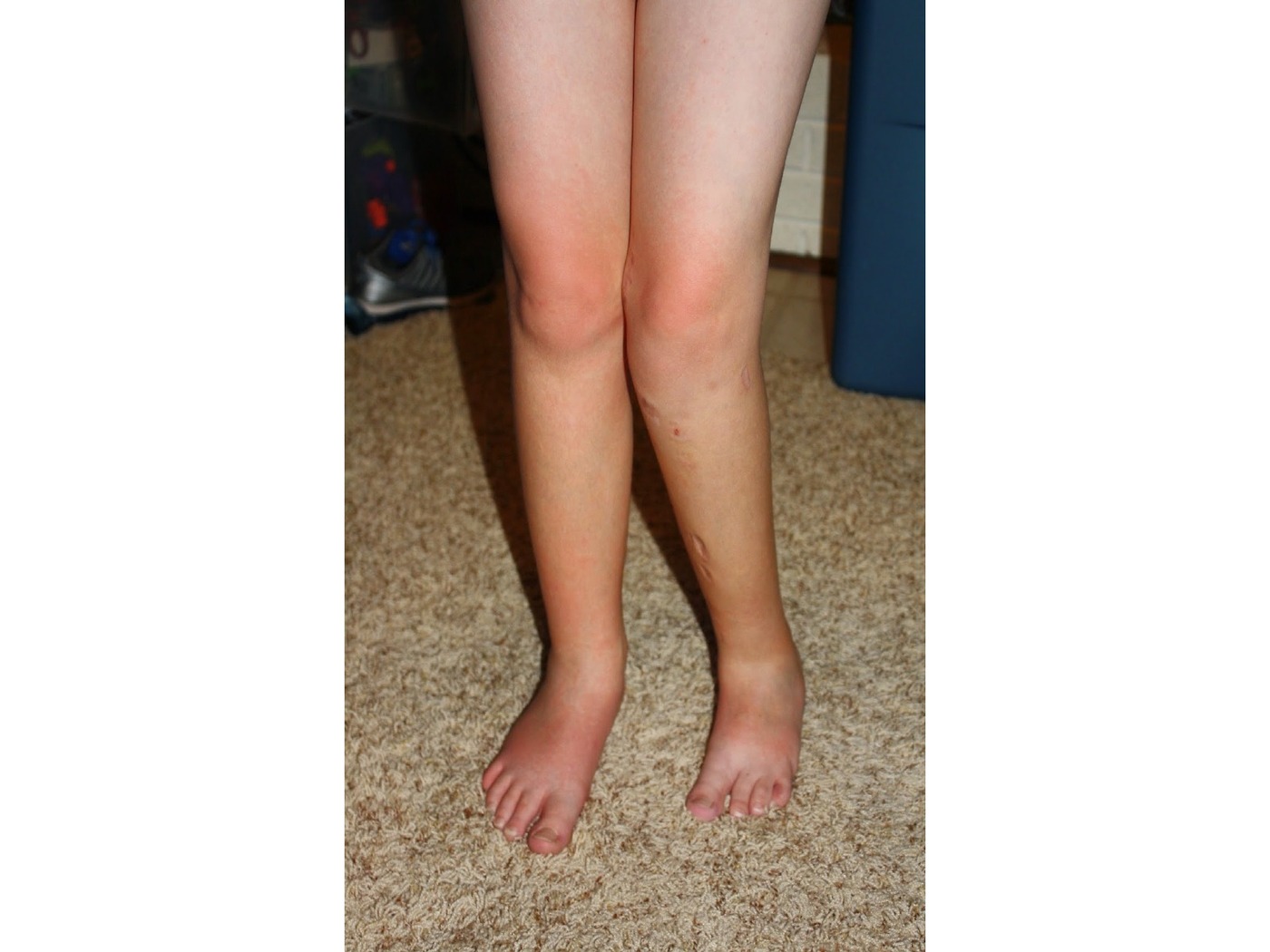
•Clinical photo showing patient standing
•Abnormalities seen over left lower limb
•LLD: tibia shorter
•Knee: valgus +/- patella subluxed
•Tibia: AM bowing, anterior scars over tibia (small, longitudinal, ?ext fix), lateral and medial scars over proximal tibia and distal femur (?previous epiphysiodesis)
•Ankle: Valgus
•Foot: Missing 5th toe, hypoplastic foot and lateral rays
•Suspect patient has fibular hemimelia (or fibular deficiency)
Differentials include trauma, tumour and infection (unlikely)
Start with history and physical examination
•To establish diagnosis, rule out associated problems, delineate current symptoms, also need to know previous treatment
PE to look for other associated abnormalities:
•LLD
•Hip: PFFD, coxa vara
•Knee: hypoplastic lateral femoral condyle, genu valgus, patella instability, ACL deficiency
•Tibia: Anteromedial bowing, multiple surgical scars
•F&A: ankle instability secondary to ball and socket ankle, talipes equinovalgus, tarsal coalition, missing lateral toes
•Scannogram
Radiographic classification by Achterman
Management
•Treatment Goal is to provide a plantigrade foot, stable ankle, and achieve equality of leg length, and to correct the tibial deformity to allow for more efficient gait
•If LLD >20cm at maturity, foot to level of mid tibia or femur <50% of other leg → ablation
•I will use Birch classification to guide my management
•Depends on whether the 1. foot is functional or not, and 2. degree of LLD
If LLD reconstructable, foot functional:
•Foot functional =>3rays and ankle stable, LLD mild = shoe raise and brace
•Foot functional =>3rays and ankle stable, LLD <5cm = contralateral epiphysiodesis
•Foot functional =>3rays and ankle stable, LLD >5cm but <30% = ipsilateral tibial lengthening
•LLD >30% = Amputation (or multiple lengthening)
•Foot non function foot / ankle unreconstructable / LLD significant
•Syme amputation if UL function ok
•Consider multiple reconstruction if poor UL function
•RECONSTRUCTION
Components:
•1. LLD
•2. Tibial bow correction
•3. Knee recon
•4. Ankle recon (soft tissue: peroneal longus release, bone: mild - MM epiphysiodesis, moderate - supramalleolar osteotomy, severe – Gruca recon (oblique sliding osteotomy → new LM)
•fibular hemimelia
•Most common congenital long bone deficiency
•Post axial longitudinal def
•Swanson: failure of formation, problem with AER, occurs in 1st trimester
Classified by Achterman & Kalamchi (based on amount of fibular present)
| Type / group | Definition / note |
|---|---|
| Type 1A | A portion of fibula remains present but proximal fibular epiphysis is distal to level of proximal tibial physis while distal fibula physis is proximal to the tibial plafond |
| Type 1B | partial absence (30-50% length), distal unable to support ankle joint |
| Type 2 | absent fibula |
Normal length of fibula:
•Proximal fibular epiphysis → proximal tibial physis
•Distal fibular physis → distal tibial plafond
•Treatment depends on the severity of LLD at maturity and stability of ankle joint
•if LLD at maturity > 30% or unstable ankle joint → symes amputation (88% satisfactory vs lengthening ~50%)