Clean FRCS notes with inline figures
Blounts disease
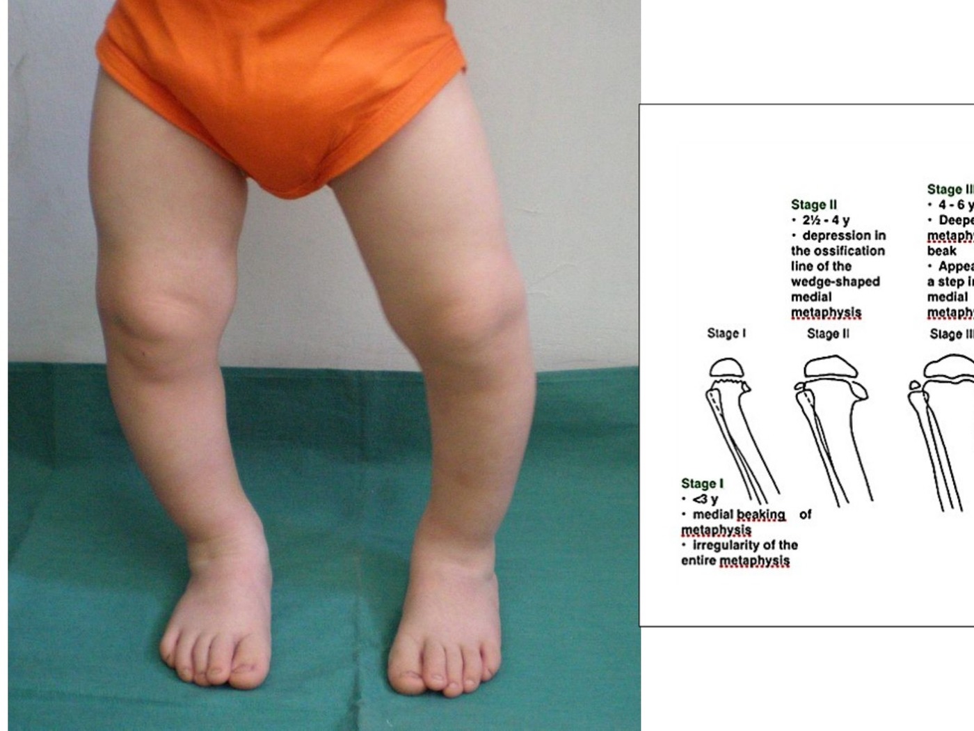
•Clinical photo showing bilateral genu varum
•Asymmetrical on both sides
•No scars, some internal rotation/ intoeing of left LL (pathognomonic for blounts)
•Likely pathological
•Can be physiological depending on AGE (Salenius curve <2 varus can be physiological)
•Causes
•Physiological, developmental, congenital (dysplasia)
•Inflammatory, metabolic, neuromuscular
•Infection, trauma, tumour
•Bilateral: metabolic, neoplastic, neuromuscular, dysplasia, developmental (DDNNM)
•Unilateral: infection, tumor (HME, Focal fibrocartilagenous dysplasia), trauma, Blount’s, iatrogenic, tibial hemimelia
History
•Onset, progression, other deformities
•Look for causes: Hx suggesting trauma, infection, tumour, Risk factors for rickets: diet
Risk factors for Blounts: early walking, obesity
Examination
•Look for causes: rickets (caput quadratum, ricketic rosary, Harrison sulcus, catback kyphosis, short stature), Dysmorphic features, CP
•Screen: Height; Body; proportion; Symmetry
•Gait: any varus thrusting
•LLD
•Check ROM, collaterals
•Measure TFA, intermalleolar distance
•Rotational profile
Systems examination: hip, spine and feet
•*Cover up test
•Normal: valgus
•Blounts: neutral / varus (indicate varus alignment at proximal tibia or knee)
XR + scannogram
•TFA, LDFA, MPTA
•Mechanical axis deviation (zoning)
•Physeal abnormalities
•Medial slope angle (angle between medial and lateral physis)
•Drennan angle (metaphyseal diaphyseal angle)
•LLD if any
•Compensatory distal femoral valgus
•Tibial-femoral subluxation
XR features of Blounts: (5)
•Varus angulation at epiphyseal – metaphyseal junction
•Widened and irregular medial physis
•Medially sloped epiphysis
•Beaking of medial metaphysis
•Lateral subluxation of tibia
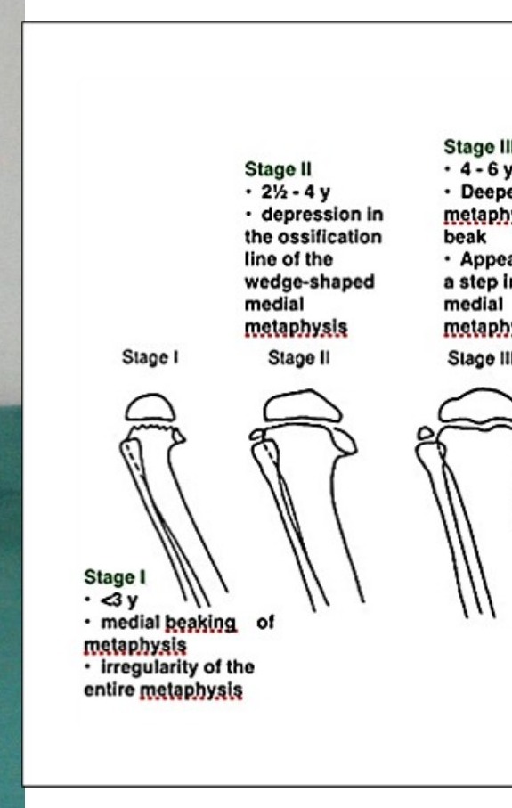
Langenskiold classification (age and XR findings)
•Beak, wedge, step, enlarge/fill, double, fused
•Bloods for CaPO4, Vit D, PTH ALP
•MRI to assess physeal bar
•+/- CT see rotation
•Aim of treatment: restore mechanical axis to unload medial physis and prevent further progression (Heuter Volkman law)
Problem list in Blount’s disease
•Varus angular deformity
•Medial physeal bar/osteochondrosis
•Depressed joint surface
•Tibial internal torsional deformity
FOR INFANTILE BLOUNTS:
•Principle: depends on patient’s age and severity
•Age cut off 4 yo
•Severity either by Langenskiold stage (III,IV, V, VI) or the Drennan angle (<9 likely physiological, >11 Blounts, >16 need OT)
•Clinical: varus thrust
OT indication:
•>4
•<4yo stage not I/II
•Drennan angle >16
| Type / group | Definition / note |
|---|---|
| I | II brace |
•KAFO with genu varus strap – provides valgus force in conjunction with a medial upright, with droplocks to increase corrective force during weight bearing
•FU with XR to look of medial physis reconstitution
•If failed to response in 1 year with progression → surgery
•III or above – brace likely not successful, gold standard:
Proximal tibial valgus osteotomy aiming for overcorrection
•+/- Physeal bar excision
•Hemiepiphysiodesis
•Hemiplateau elevation
•Correct IR
Other treatment options:
•Guided growth, with tension band plate and screws
•Possible in earlier stages
FOR ADOLESCENT FORM OF BLOUNTS:
•Less deformity, unilateral, africoamerican descent
•Tx of adolescent form Blount’s (onset > 10yo) is different, which solely depends on the varus angle >10 deg.
•Use CT/MRI to look if the physis is closed or not
•Physis open: lateral tibial epiphysiodesis (either permanent or temporary)
•Physis closed: osteotomy w/out any overcorrection either by a HTO or EF/Ilizarov method/ TSF+/- prophylactic fasciotomy
What is Blount’s disease?
•Dyschondrosis of medial proximal tibial physis leading to progressive pathological genu varum centred at the tibia
•Related to mechanical overload in genetically susceptible individuals
•Excessive medial pressure produces an osteochondrosis of the medial proximal tibial physis and epiphysis +/- progress to physeal bar
•(Heuter Volkman’s law: increased stress = less growth in physis)
Pathophysiology explained by HV principle
•Increase loading to proximal tibial PM physis, growth suppression and irreversible osteochondrosis + bone bridging at medial proximal tibial physis. Also marked metaphyseal fragmentation and resultant progressive tibial vara.
•Vs physiological varus
•Progressive
•More abrupt (Drennan angle >11)
•Physeal abnormality
•Asymmetrical flaring
Risk factors for recurrence
•Age > 5
•Body weight > 95 percentile
•Medial physeal slope > 60 degree
•Langenskiöld stage IV or above
•Drawback of Langenskiold staging
•All stages can occur earlier than described age
•Stage II and III could progress to VI despite OT (i.e. OT does not change the natural history)
•Single osteotomy gives good results only in <4 yo but not <8 yo
•Only include white population instead of more severe Afro-American population