Clean FRCS notes with inline figures
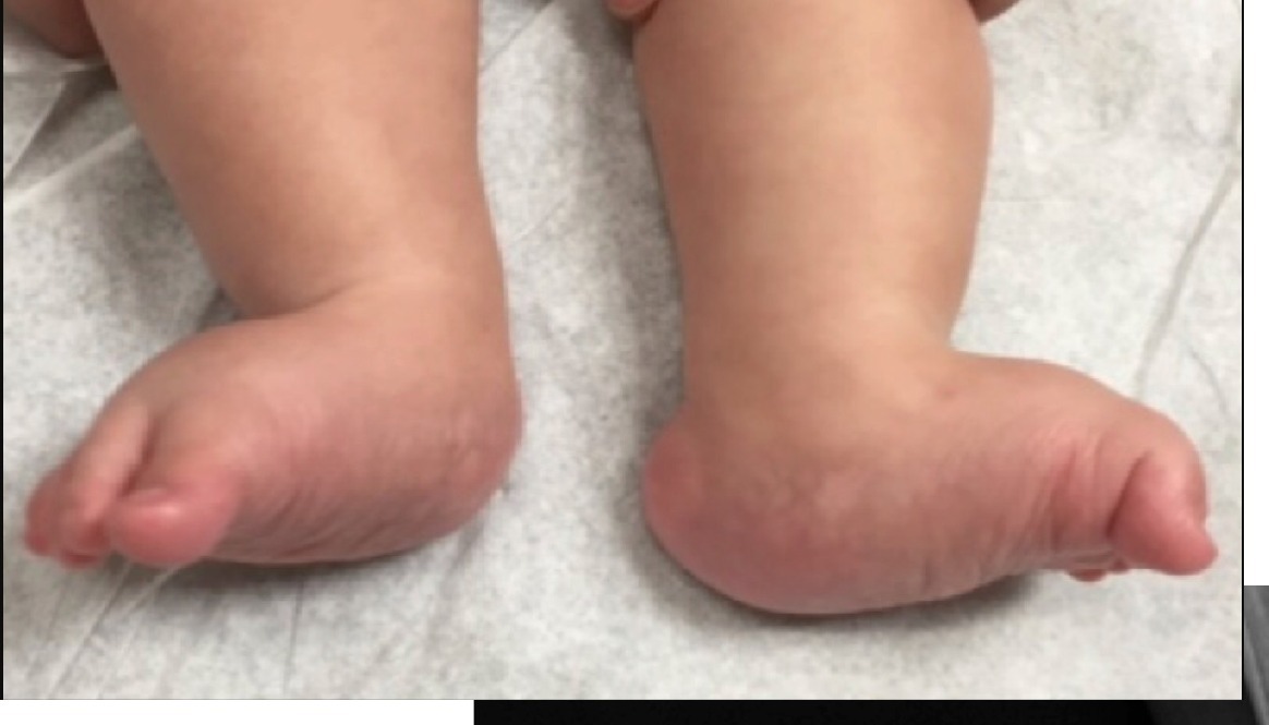
Clinical photograph
•Midfoot: Rocker bottom deformity
•Equinus hind foot
•Dorsiflexion and abduction of forefoot
Rigid flatfoot ddx:
•Tarsal coalition
•CVT
•Accessory navicular
•+/- stage 3 or 4 PTTD in adults
Radiographic description
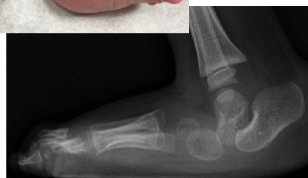
•Vertically pointed talus
•Long axis of talus passes below the 1st ray axis (NOT navicular as it is not ossified yet)/ TAMBA (Talus axis MT bone angle)
•Increase in talocalcaneal angle
•Take plantarflexion lateral XR to distinguish between CVT and COT
Diagnosis – CVT
Differential diagnosis
Congenital oblique talus (plantarflexion lateral XR)
•Calcaneovalgus (Calcaneus instead of equinus, break at hindfoot instead)
•Tarsal coalition
•Paralytic pes planus
How will you assess this child?
•Systemically screen for associations
•50% +ve neuromusclular disease (spina bifida), syndromal (arthrogryposis, Larsen syndrome), maltreated CTEV, look for DDH
•Genetic: 20% +ve family Hx
•Locally confirm rigid deformity
•Rocker bottom foot
•Hind foot: equinovalgus deformity, tight TA and peroneus
•Chopart joint: Dorsiflexion with navicular dorsal dislocation
•Forefoot: abduction and dorsiflexion (EDL/ EHL, tib ant contractures)
•Prominent talar head at plantar region
•Check LL neurology
•“peg leg” gait
Xray:
•Lat: Vertically pointed talus, long axis of talus passes below the 1st ray axis (Dorsally dislocated navicular, navicular ossify at 3 y.o.)
•AP: Increase in talocalcaneal angle >40
•Differentiate between CVT and COT: try to reduce TNJ in plantarflexion view
Management
•r/o association
•r/o oblique talus (planter flexed Xray, if reducible → oblique talus)
•Counselling: May try reverse Ponseti but need to tell parents majority eventually need OT
•Preop stretching of dorsolateral structure + reverse ponsetti x 3 mths (identify the talar head, push forefoot in plantar medial direction)
•After reduction perform talonavicular pinning (MIS) + release peroneus and extensors +/- lengthening +/- spring ligament recon + percutaneous achilles tenotomy 6 to 12 months after casting
•JBJS (Am) 2006 Dobbs: 11 patients treated with stretching + reverse ponseti + percutaneous pinning and achilles tendon tenotomy, good result in appearance, foot function and Xray at minimum 2 years
•If present late: talus + navicular excision and lateral column lengthening
•Salvage: triple arthrodesis
•Vertical talus
•Irreducible dorsal dislocation of the navicular on the talus producing a rigid flatfoot deformity
What is the pathoanatomy?
•Soft tissue: Tight Achilles, displaced PT and peroneal both function as dorsiflexor, attenuated spring ligament
•Bony: dorsally dislocated navicular
•COT
•No need serial casting
•Treated with TA release