FRCS Paediatric Ortho — improved notes
FRCS Paediatric Ortho — visual inline notes

036. Painful flatfoot onset in adolescence: Tarsal coalition

visual-inline rebuild3 extracted figuresspeaker notes XML87 study lines8 Q&A

Individual slide images have been cropped from the rendered slide and inserted beside the matching note section. The full slide remains below only for layout/audit.

Clean FRCS notes with inline figures

Painful flatfoot onset in adolescence: Tarsal coalition

Clinical photograph
Clinical photograph - tarsal coalition clinical photograph
Fig 1. Clinical photograph - tarsal coalition clinical photographmiddle left
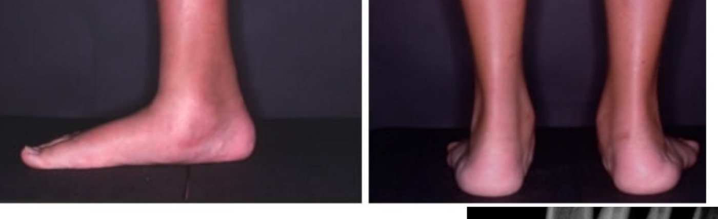
•Bilateral flatfoot with reduced medial foot arch
•Medial bulge → middle facet coalition
•From the back, hindfoot valgus with too many toe sign positive on right side

Diagnosis: pes planus

•Check/perform differentiate if it is flexible or rigid
•Ddx for painful flatfoot in child
•Tarsal coalition
•Accessory navicular
•Painful flexible flatfoot (tight Achilles)
•JIA

Causes of flatfoot in general:

•Unilateral vs bilateral vs rigid
•IDNNM cause

Assessment:

•Hx: Onset of symptoms, site of pain, trauma, footwear

Examination

•Exclude obvious syndromic or neurological causes
•Deformity (hindfoot valgus, forefoot abduction, rigid flatfeet)
•Tenderness (microfracture of coalition interface, ossification of previously fibrous or cartilaginous coalition, chondral injury, peroneal spasm, subfibular impingement, increase stress in other joints → hindfoot joint degeneration) (CN lateral pain at sinus tarsi, TC medial pain)
•Painful inversion against resistance (peroneal spasm)
•Limited subtalar movement. Reverse Coleman block to assess subtalar rigidity
•PTT function
•Examine contralateral side (50% bilateral)
•General exam: Associated with fibular hemimelia/ PFFD, carpal coalition, Apert syndrome
•CN vs TC coalition
•CN
•- More common, present earlier
•- Less stiff
•- lateral side pain
•- More responsive to resection

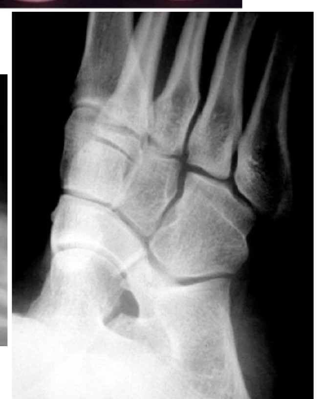
- Anteater nose sign on oblique XR
Radiograph / imaging - tarsal coalition radiograph
Fig 2. Radiograph / imaging - tarsal coalition radiographtop left
Radiograph / imaging - tarsal coalition radiograph
Fig 3. Radiograph / imaging - tarsal coalition radiographmiddle centre
•TC
•- Less common
•- Present late
•- Medial side pain
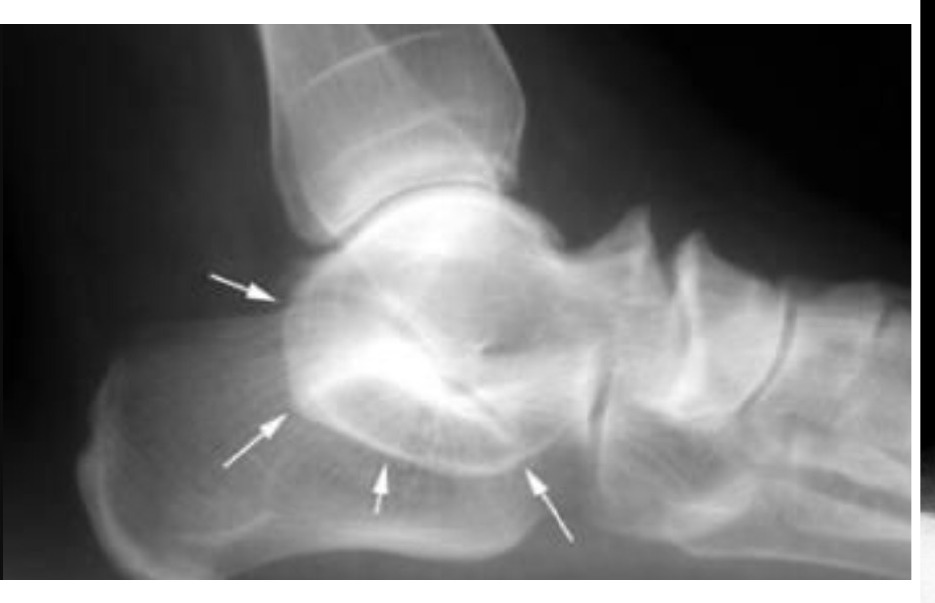
•- C sign and anterior beak on lateral XR, coalition in harris view

Xray:

•CN: oblique view showed anteater sign
•TC: lateral view showed C sign= subtalar and middle facet coalition, talus neck periosteal traction spur. Harris view may also show coalition
•CT/ MRI to look for size and location of bony/ fibrous connection, r/o additional coalitions (5%)

Treatment:

•Conservative (if rigid flatfoot, arch support/ orthosis mold at current position to prevent deterioration eg UCBL) (walking cast boot) (temporary immobilization with casting)
•OT: Resection of coalition vs fusion

Myerson:

•Traditionally 50% is regarded as a contraindication for resection. Most important aspect of treatment is to realign the foot and ankle regardless of fusion or resection
•CN: excision via Olliers approach/sinus tarsi approach, EDB interposition +/- correct deformity and TA release
•TC: bony union > 50% → fusion, <50% → excision + interposition (split FHL graft, fat)
•Medial incision between FDL and PT
•Salvage: triple arthrodesis + deformity correction in rigid FF for older patient + OA

Indications for fusion:

•1. Malalignment
•2. >50%
•3. Degen
•4. Older

•AD – high level of penetrance
•NOG (Noggin) gene
•Bilateral in 50%
•Failure of mesenchymal segmentation

Ddx for flatfoot:

•Unilateral TDNNM
•Post traumatic
•Degenerative
•Charcot (neuroinflammatory)
•Neuromuscular (polio)
•Bilateral IDNNM
•Ligamentous laxity
•RA (inflammatory)
•Neuroinflammatory
•Neuromuscular (spastic diplegic, spina bifida)
•Both
•Idiopathic
•PTTD
•Congenital vertical talus
•Tarsal coalition

What does peroneal spasm mean?

•Peroneal spasm is a protective mechanism to reduce pain
•If no peroneal spasm, you know subtalar joint is okay (Lui)

Canadian army foot study

•1/5 have flat foot
•67% asx
•25% sx with flexible flat foot + tight TA
•8% sx with rigid (accessory navicular, tarsal coalition, congenital vertical talus)
•Wagner JBJS 1989
•Flexible flatfoot randomized to control, corrective shoes, Helfet heel cups, UCBL – improvement not related to orthosis. Increase laxity, more improvement.

Direct Q&A

Q1 What clinical signs are shown for Painful flatfoot onset in adolescence: Tarsal coalition?

Bilateral flatfoot with reduced medial foot arch

•Medial bulge → middle facet coalition
•From the back, hindfoot valgus with too many toe sign positive on right side
Q2 What assessment points are listed for Painful flatfoot onset in adolescence: Tarsal coalition?

Hx: Onset of symptoms, site of pain, trauma, footwear

Q3 What examination findings should be assessed in Painful flatfoot onset in adolescence: Tarsal coalition?

Exclude obvious syndromic or neurological causes

•Deformity (hindfoot valgus, forefoot abduction, rigid flatfeet)
•Tenderness (microfracture of coalition interface, ossification of previously fibrous or cartilaginous coalition, chondral injury, peroneal spasm, subfibular impingement, increase stress in other joints → hindfoot joint degeneration) (CN lateral pain at sinus tarsi, TC medial pain)
•Painful inversion against resistance (peroneal spasm)
•Limited subtalar movement. Reverse Coleman block to assess subtalar rigidity
•PTT function
•Examine contralateral side (50% bilateral)
•General exam: Associated with fibular hemimelia/ PFFD, carpal coalition, Apert syndrome
•CN vs TC coalition
•CN
•- More common, present earlier
•- Less stiff
•- lateral side pain
•- More responsive to resection
Q4 What are the radiographic findings for Painful flatfoot onset in adolescence: Tarsal coalition?

TC

•- Less common
•- Present late
•- Medial side pain
•- C sign and anterior beak on lateral XR, coalition in harris view
Q5 How is Painful flatfoot onset in adolescence: Tarsal coalition managed?
•Conservative (if rigid flatfoot, arch support/ orthosis mold at current position to prevent deterioration eg UCBL) (walking cast boot) (temporary immobilization with casting)
•OT: Resection of coalition vs fusion
Q6 What are the indications listed for Painful flatfoot onset in adolescence: Tarsal coalition?
•1. Malalignment
•2. >50%
•3. Degen
•4. Older
Q7 What does peroneal spasm mean?

Peroneal spasm is a protective mechanism to reduce pain

•If no peroneal spasm, you know subtalar joint is okay (Lui)
Q8 What evidence or study finding is cited for Painful flatfoot onset in adolescence: Tarsal coalition?

1/5 have flat foot

•67% asx
•25% sx with flexible flat foot + tight TA
•8% sx with rigid (accessory navicular, tarsal coalition, congenital vertical talus)
•Wagner JBJS 1989
•Flexible flatfoot randomized to control, corrective shoes, Helfet heel cups, UCBL – improvement not related to orthosis. Increase laxity, more improvement.

Raw speaker-note hierarchy audit

#lvlmarLindentlogicaloriginal note line
10000Painful flatfoot onset in adolescence: Tarsal coalition
20NoneNone0Describe clinical photo:
30171450-1714501Bilateral flatfoot with reduced medial foot arch
40171450-1714501Medial bulge → middle facet coalition
50171450-1714501From the back, hindfoot valgus with too many toe sign positive on right side
60NoneNone0Diagnosis: pes planus
70NoneNone0Need to differentiate if it is flexible or rigid
80NoneNone0Ddx for painful flatfoot in child
90171450-1714501Tarsal coalition
100171450-1714501Accessory navicular
110171450-1714501Painful flexible flatfoot (tight Achilles)
120171450-1714501JIA
130NoneNone0Causes of flatfoot in general:
140NoneNone0Unilateral vs bilateral vs rigid
150NoneNone0IDNNM cause
160NoneNone0Assessment:
170NoneNone0Hx: Onset of symptoms, site of pain, trauma, footwear
180NoneNone0PE:
191171450-1714501Exclude obvious syndromic or neurological causes
201171450-1714501Deformity (hindfoot valgus, forefoot abduction, rigid flatfeet)
211171450-1714501Tenderness (microfracture of coalition interface, ossification of previously fibrous or cartilaginous coalition, chondral injury, peroneal spasm, subfibular impingement, increase stress in other joints → hindfoot joint degeneration) (CN lateral pain at sinus tarsi, TC medial pain)
221171450-1714501Painful inversion against resistance (peroneal spasm)
231171450-1714501Limited subtalar movement. Reverse Coleman block to assess subtalar rigidity
240171450-1714501PTT function
250171450-1714501Examine contralateral side (50% bilateral)
260171450-1714501General exam: Associated with fibular hemimelia/ PFFD, carpal coalition, Apert syndrome
270000CN vs TC coalition
280000CN
290000- More common, present earlier
300000- Less stiff
310000- lateral side pain
320000- More responsive to resection
330000- Anteater nose sign on oblique XR
340000TC
350000- Less common
360000- Present late
370000- Medial side pain
380000- C sign and anterior beak on lateral XR, coalition in harris view
390NoneNone0Xray:
400171450-1714501CN: oblique view showed anteater sign
410171450-1714501TC: lateral view showed C sign= subtalar and middle facet coalition, talus neck periosteal traction spur. Harris view may also show coalition
420000CT/ MRI to look for size and location of bony/ fibrous connection, r/o additional coalitions (5%)
430NoneNone0Treatment:
440171450-1714501Conservative (if rigid flatfoot, arch support/ orthosis mold at current position to prevent deterioration eg UCBL) (walking cast boot) (temporary immobilization with casting)
450171450-1714501OT: Resection of coalition vs fusion
46145720001Myerson:
47145720001Traditionally 50% is regarded as a contraindication for resection. Most important aspect of treatment is to realign the foot and ankle regardless of fusion or resection
481628650-1714502CN: excision via Olliers approach/sinus tarsi approach, EDB interposition +/- correct deformity and TA release
491628650-1714502TC: bony union > 50% → fusion, <50% → excision + interposition (split FHL graft, fat)
5021085850-1714503Medial incision between FDL and PT
511628650-1714502Salvage: triple arthrodesis + deformity correction in rigid FF for older patient + OA
520000Indications for fusion:
5300001. Malalignment
5400002. >50%
550NoneNone03. Degen
560NoneNone04. Older
570NoneNone0=============================
580NoneNone0AD – high level of penetrance
590NoneNone0NOG (Noggin) gene
600NoneNone0Bilateral in 50%
610NoneNone0Failure of mesenchymal segmentation
620NoneNone0Ddx for flatfoot:
630NoneNone0Unilateral TDNNM
641NoneNone1Post traumatic
651NoneNone1Degenerative
661NoneNone1Charcot (neuroinflammatory)
671NoneNone1Neuromuscular (polio)
680NoneNone0Bilateral IDNNM
691NoneNone1Ligamentous laxity
701NoneNone1RA (inflammatory)
711NoneNone1Degenerative
721NoneNone1Neuroinflammatory
731NoneNone1Neuromuscular (spastic diplegic, spina bifida)
740NoneNone0Both
751NoneNone1Idiopathic
761NoneNone1PTTD
771NoneNone1Congenital vertical talus
781NoneNone1Tarsal coalition
790NoneNone0What does peroneal spasm mean?
800NoneNone0Peroneal spasm is a protective mechanism to reduce pain
810NoneNone0If no peroneal spasm, you know subtalar joint is okay (Lui)
820NoneNone0Canadian army foot study
831NoneNone11/5 have flat foot
841NoneNone167% asx
851NoneNone125% sx with flexible flat foot + tight TA
861NoneNone18% sx with rigid (accessory navicular, tarsal coalition, congenital vertical talus)
870NoneNone0Wagner JBJS 1989
880NoneNone0Flexible flatfoot randomized to control, corrective shoes, Helfet heel cups, UCBL – improvement not related to orthosis. Increase laxity, more improvement.

Improved semantic hierarchy used for the study notes

#semantic levelsection?rewritten line
10Painful flatfoot onset in adolescence: Tarsal coalition
20yesClinical photograph
31Bilateral flatfoot with reduced medial foot arch
41Medial bulge → middle facet coalition
51From the back, hindfoot valgus with too many toe sign positive on right side
60yesDiagnosis: pes planus
71Check/perform differentiate if it is flexible or rigid
81Ddx for painful flatfoot in child
91Tarsal coalition
101Accessory navicular
111Painful flexible flatfoot (tight Achilles)
121JIA
130yesCauses of flatfoot in general:
141Unilateral vs bilateral vs rigid
151IDNNM cause
160yesAssessment:
171Hx: Onset of symptoms, site of pain, trauma, footwear
180yesExamination
191Exclude obvious syndromic or neurological causes
201Deformity (hindfoot valgus, forefoot abduction, rigid flatfeet)
211Tenderness (microfracture of coalition interface, ossification of previously fibrous or cartilaginous coalition, chondral injury, peroneal spasm, subfibular impingement, increase stress in other joints → hindfoot joint degeneration) (CN lateral pain at sinus tarsi, TC medial pain)
221Painful inversion against resistance (peroneal spasm)
231Limited subtalar movement. Reverse Coleman block to assess subtalar rigidity
241PTT function
251Examine contralateral side (50% bilateral)
261General exam: Associated with fibular hemimelia/ PFFD, carpal coalition, Apert syndrome
271CN vs TC coalition
281CN
291- More common, present earlier
301- Less stiff
311- lateral side pain
321- More responsive to resection
330yes- Anteater nose sign on oblique XR
341TC
351- Less common
361- Present late
371- Medial side pain
381- C sign and anterior beak on lateral XR, coalition in harris view
390yesXray:
401CN: oblique view showed anteater sign
411TC: lateral view showed C sign= subtalar and middle facet coalition, talus neck periosteal traction spur. Harris view may also show coalition
421CT/ MRI to look for size and location of bony/ fibrous connection, r/o additional coalitions (5%)
430yesTreatment:
441Conservative (if rigid flatfoot, arch support/ orthosis mold at current position to prevent deterioration eg UCBL) (walking cast boot) (temporary immobilization with casting)
451OT: Resection of coalition vs fusion
460yesMyerson:
471Traditionally 50% is regarded as a contraindication for resection. Most important aspect of treatment is to realign the foot and ankle regardless of fusion or resection
482CN: excision via Olliers approach/sinus tarsi approach, EDB interposition +/- correct deformity and TA release
492TC: bony union > 50% → fusion, <50% → excision + interposition (split FHL graft, fat)
503Medial incision between FDL and PT
512Salvage: triple arthrodesis + deformity correction in rigid FF for older patient + OA
520yesIndications for fusion:
5311. Malalignment
5412. >50%
5513. Degen
5614. Older
570——
581AD – high level of penetrance
591NOG (Noggin) gene
601Bilateral in 50%
611Failure of mesenchymal segmentation
620yesDdx for flatfoot:
631Unilateral TDNNM
641Post traumatic
651Degenerative
661Charcot (neuroinflammatory)
671Neuromuscular (polio)
681Bilateral IDNNM
691Ligamentous laxity
701RA (inflammatory)
711Neuroinflammatory
721Neuromuscular (spastic diplegic, spina bifida)
731Both
741Idiopathic
751PTTD
761Congenital vertical talus
771Tarsal coalition
780yesWhat does peroneal spasm mean?
791Peroneal spasm is a protective mechanism to reduce pain
801If no peroneal spasm, you know subtalar joint is okay (Lui)
810yesCanadian army foot study
8211/5 have flat foot
83167% asx
84125% sx with flexible flat foot + tight TA
8518% sx with rigid (accessory navicular, tarsal coalition, congenital vertical talus)
861Wagner JBJS 1989
871Flexible flatfoot randomized to control, corrective shoes, Helfet heel cups, UCBL – improvement not related to orthosis. Increase laxity, more improvement.