Clean FRCS notes with inline figures
Clinical photo showing paed patient with bilateral feet deformity
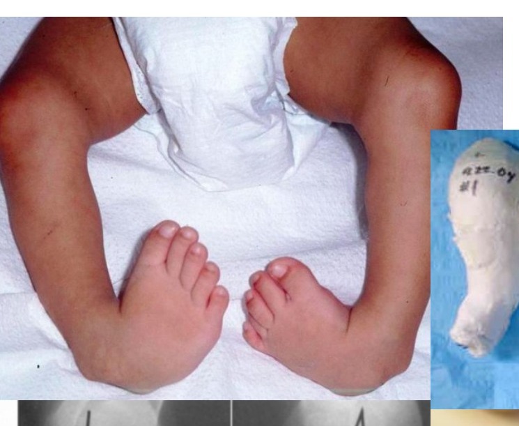
•Hindfoot in varus and equinus, midfoot in cavus and forefoot in adduction + pronated position
•Bilateral clubfoot
•Congenital talipes equinovarus (CTEV)
•Locally assess correctability (?postural), assess the other side (50% bilateral)
•Systemically assess associated problem
•Packaging syndrome (torticollis, DDH, knee dislocation)
•Rule out “syndromic” club foot
•NM disease (CP)
•Spinal dysraphism
•Other teratogenic /syndrome (arthrogryposis, SED, constriction band, Larsen, Pierre Robin, Prune Belly)
•XR controversial: look for parallelism of talus and calcaneum
How to counsel parents?
•Familial occurrence in 25%. Affected child have 2.5-6.5% chance of another child with clubfoot
•[PITX gene, chromosome 2 deletion (Common genetic pathway with CVT)]
•Treatment – Ponseti casting, success rate >90%, 70% need PETA, 30% need tib ant transfer for dynamic supination at age 4-5
•Affected foot will be smaller and shorter regardless of treatment
•If it is a structural deformity, I would offer non-operative management with serial casting by Ponseti method
•It is based on the viscoelastic property of the soft tissue with the principle of stress relaxation & creep
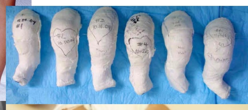
•Start at 1 week, toe to groin cast with POP with knee in 90deg. Cast has to be snug but not tight, thin layer of velband only, followed by well molded cast
•Change cast: bath the cast and unwind cast
Correction sequence CAVE
•Cavus (DF 1st ray, realign plantarflexed 1st MT with other MT, realign forefoot to hindfoot by supination) [warn family that deformity may look worse]
•Adductus (abduct forefoot and ER at midfoot level using the uncovered head of talus as fulcrum, locate talus as just anterior to fibular)
•Varus (will be corrected with #2)
•Equinus (pull calcaneal tuberosity distally while pushing most anterior part of calcaneus up, when abduction 70deg achieved, able to palpate the anterior process of the calcaneus as it abducts out from beneath the talus, neutral or slight heel valgus)
•Check/perform get the assistant to hold the big toe and talar head for the entire duration
•Weekly change cast, usually need 4-5 casts
•Chart progress with Pirani score (high score = more cast needed and more chance of relapse)
•Midfoot: medial skin crease, curve lateral border, talar head coverage
•Hindfoot: posterior skin crease, absent heel, equinus rigidity
•70% patient require TA lengthening (NA, incision medial to TA, transverse cut towards lateral), then to another cast for 3/52 at 30 deg ankle DF
•(indicated if everything corrected but DF still <10deg)
PETA:
•From posteromedial, just next to Achilles, towards lateral
•Then change blade 90deg and dorsiflex → feel pop
•Then to DB boots full day x 4 months (usually until 6month) then at night & naps 4 years
•30% patient has dynamic supination during swing phase, require operation x transfer of TA from medial cuneiform to cuboid / lateral cuneiform
•(@ 4-5 yo for ossification)
•If failed conservative management (foot not plantigrade by 6m)
OT timing controversial, weight bearing to avoid recurrence?
•Simmons – size more important >=8cm
•Posteromedial release with Cincinati incision, a la cart approach
•Do not release
•1. Deep talocalcaneal interosseous ligament (risk of lateral translation of calcaneus)
•2. Deep deltoid ligament (valgus deformity)
•3. Posterior tibio-fibular ligament
Describe the pathoanatomy of CTEV
•Primary deformity is centered at the talus, which has a medially and plantar deviated neck. The rest of the acetabulum pedis (calcaneus, navicular, cuboid) then rotates around the talus and becomes adducted and inverted.
•Secondary soft tissue contracture (plantarflexor + invertor shortening; contracted CFL, deltoid, long + short plantar, spring, plantar aponeurosis)
Causes:
•Majority are idiopathic
•Neuropathic theory
•Myopathic theory
•Genetic (PITX gene)
•Mechanical molding theory
•Multifactorial
Would you XR every clubfoot?
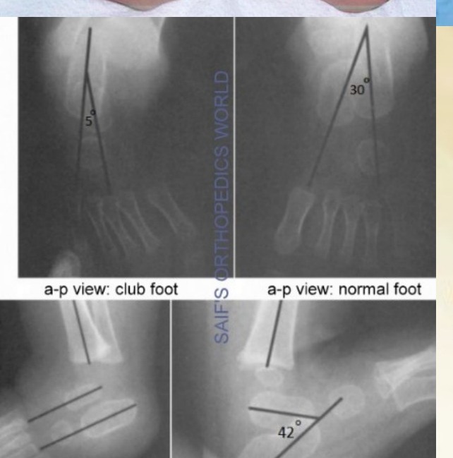
Xray is controversial
•Difficult to position
•Only calcaneum + talus + MT ossified (Navicular ossify at 4yo)
•Ossification center does not represent true shape
•**Double parallelism of talus and calcaneum
•Dorsiflexion lateral (Turco view)
•Hindfoot parallelism
•Talocalcaneal angle < 35°
•AP view
•Kite angle (talocalcaneal) <20
•talus-first metatarsal angle –ve
Complications of casting
•Rockerbottom foot (2-5%)
•Flat heel pad
•Crowded toes
•Pressure sores
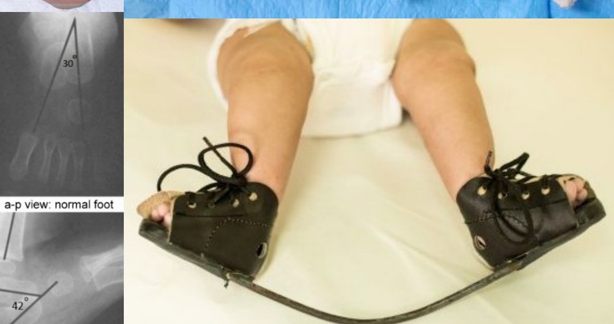
•Denis Brown Boots
•For patients with club foot after serial casting
•BOOTS
•Strap for 3 point fixation
•Forefoot – open shoe box
•Midfoot – scaphoid and intermetatarsal pad, straight medial lass to keep the foot abducted
•Hindfoot – reverse Thomas heel, stiff heel support for ML stability
•BAR
•Convex away to keep ankle dorsiflexion 5-10deg
•Shoulder width
•ER 70/40, both sides 45 (sum 90-100) to keep hindfoot in valgus
•Regime
•4m full time, till 4y night time