Clean FRCS notes with inline figures
•Physeal bar
AP Xray of knee of skeletally immature patient
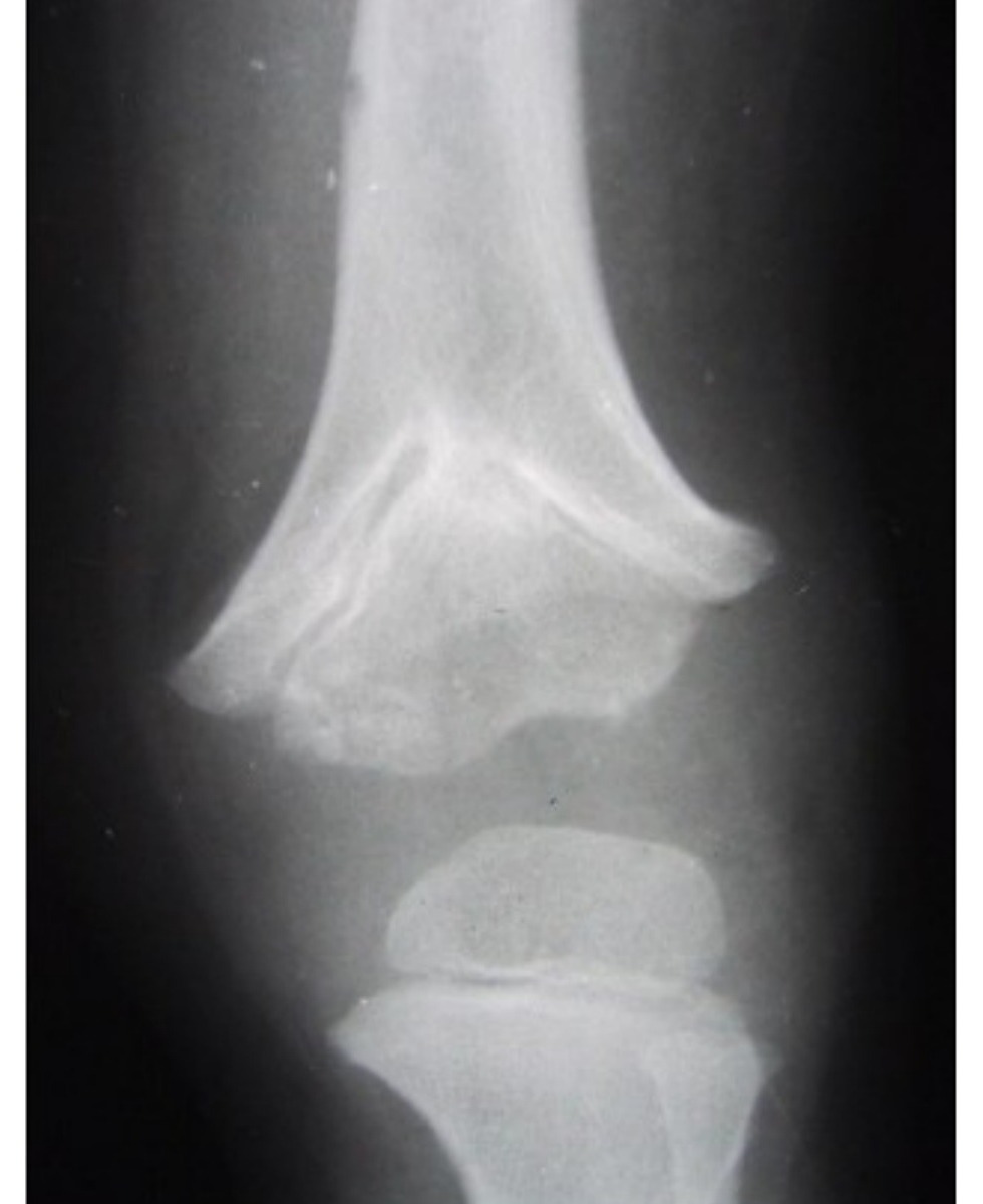
•V shaped physis of distal femoral physis, suspected connection between metaphyseal and epiphyseal area in middle (cross trabeculation)
•Suspected physeal arrest
•Would like to have long film involving contralateral limb to see LLD, angular deformity
Also lateral Xray to see angular deformity
Sequelae:
•LLD, angular deformity, epiphyseal deformity
•Hx: age, causes
Examination
•Local: deformity, length, joint involvement, features suggesting cause
•Systemic: maturity
Investigations
•CT to look for bony bar, MRI to look for fibrous tissue, also to look for underlying cause
Classification:
•Extent (partial / complete)
•Site (peripheral, central, linear)
Prognosis affected by
•Cause
•Extent
•Site
•Growth potential
•Management principle: achieve equal leg length with no angular deformities, minimize complications and hospital stay
Treatment depends on
•Growth potential and projected LLD
•Deformity present: angular, LLD
•Area / location involved
•More than 2 years growth remaining
•<50% physis involved
•Langenskiold procedure: Physeal bar excision (direct/ metaphyseal window/ through osteotomy) with interposition (bone reformation retardant eg fat, cement to avoid hematoma) + marker implantation
•Physis distraction
•>50% physis involved – hemi/epiphysiodesis + future lengthening
•Less than 2 years growth remaining
•Hemiepiphysiodesis
•No growth
•Osteotomy
•FU till maturity
Prognosis:
•Traumatic: responsive to physeal bar excision as only hypertrophic zone affected
•Tumor/ infection: decrease response as all zones affected
•JPO 2009
•Factors to determine risk of physeal bar in fractures
•Initial displacement
•Mechanism of injury (high)
•Pattern SH
•Number of reduction (not more than 2)
•Residual displacement after CR
What causes growth arrest?
•Injury to germinal matrix (resting zone)
Affecting blood supply
•Bone bar formation