FRCS Paediatric Ortho — improved notes
FRCS Paediatric Ortho — visual inline notes

041. LLD

visual-inline rebuild1 extracted figuresspeaker notes XML121 study lines6 Q&A

Individual slide images have been cropped from the rendered slide and inserted beside the matching note section. The full slide remains below only for layout/audit.

Clean FRCS notes with inline figures

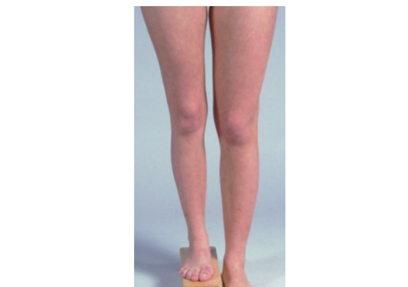
Clinical photograph

•Block test
•Right LL shorter by ~5cm
•Both femur and tibia are involved
•Foot and ankle no gross deformity
•No skin stigmata (Café au lait spots in NF in tibial pseudoarthrosis)
•Presence of all 5 toes (fibular hemimelia)

OVERALL:

•Look for deformity: LLD / angular / rotation

Cause:

•Hemihypertrophy: Vascular, NF
•Shortening: Neurological, congenital, local (physis)
•Bone age (clinical / radiological)
•Predict LLD (Shapiro + calculations)
•Management plan: conservative / epiphysiodesis / lengthening / amputation
•Cause of LLD

Long leg:

•Congenital hemihypertrophy (e.g. NF, Klippel Trenaunneay, Beckwidth Wiedemann, Proteus)
•Physeal stimulation (post traumatic)

Short leg:

•Congenital (DDH, fibular hemimelia, PFFD, calcaneovalgus, enchondromatosis)
•Neuromuscular (polio, cerebral palsy)
•Vascular (Perthes, AV fistula)
•Physis (physis bar, hyperemia): trauma, infection, tumor

Management:

•Assess cause (from femur/ tibia)
•Assess skeletal maturity (clinical, radiological)
•Calculate LLD at maturity
•Assess function of foot and ankle (joint mobility and stability)
•R/O other deformity (angular, rotational, LL contracture)
•Assess skeletal maturity
•Clinical
•Chronological age
•Tanner staging (1st sign: enlargement of testis, breast bud. Descending phase: menarche/axillary hair)
•Menarche
•Growth spurt
•Radiological
•1. Tanner Whitehouse (With reference to the morphology and fusion of physis in hand and wrist)
•2. Gruelich and Pyle atlas (DP→MC→PP→MP→ulnar→radius)
•3. Risser
•4. Elbow epiphyseal centre (Sauvergain)
•Calculation for LLD at maturity

Multiple FU:

•1 Moseley chart (need leg length of both LL and Xray hand for skeletal maturity)
•2 Arithmetic method (calculate growth inhibition and predict LLD by future growth in long leg x inhibition)
•Assumption: distal femur 9mm/yr, proximal tibia 6mm/yr, boy stop growth 16, girl 14, Shapiro type 1
•3 Green Anderson chart

Single FU:

•1 Paley multiplier method (LLD X age and sex factor)
•2 Simplistic
•Also taken into consideration Shapiro’s growth pattern
•LLD proportionally increase: Physeal arrest, PFFD – Most common, static growth inhibition
•LLD growth decelerates: NM conditions
•LLD static after a while: Overgrowth of femur fracture
•LLD static after a while then increase again: Perthes
•LLD plateau and reduce: JRA

General principles:

•Depends on the LLD at skeletal maturity
•<2cm: observe (manage with shoe raise)
•2-5cm: shorten longer limb by epiphysiodesis if physis open, shortening osteotomy if physis closed
•>5cm: lengthen short limb +/- shorten long limb
•>20cm: consider amputation
•Lengthening techniques (lengthen the short side)

Acute

•Gradual
•Distraction histiogenesis
•Gradual lengthening procedure by intramembranous ossification, use of distraction force to stimulate the formation of bone and soft tissue

Procedure:
Operative / approach image
Fig 1. Operative / approach imagetop left
•Put on exfix
•Perform low energy corticotomy at metaphyseal region (transecting 2/3 of circumference of cortex and complete by osteoclasis by rotating the proximal and distal segment in opposite direction. Preserve the periosteum, endosteum, medullary content and nutrient artery ) OR osteotomy (multiple drill holes followed by completion by osteotome) = no difference in the literature
•Rest for 5-7days
•Gradual distraction (1mm per day divided into 4 times)
•Consolidation ~2x time of lengthening, need Xray to confirm formation of at least 3 cortex
•Removal of exfix when there is closure of interzone and 2 side neocorticalisation on AP and lat films
•Maximum lengthening each time: 20%
•(Michael To tutorial: JPO 2010? % Lengthening related to % of complication, if lengthening >20% → increase complication e.g. nonunion, NV injury, infection, contracture)

Complication:

•Angular deformity
•Pin tract infection
•Nerve injury (during OT/ traction injury)
•Fracture due to early removal of ex fix
•Chondrodiastasis
•Gradual distraction of physis result in hypertrophy of cellular layers of the physis without separation - Heuter Volkman
•Unpredictable results
•initial lengthening is often followed by growth plate fusion
•Shortening techniques (shorten the long side)
•Permanent
•Open epiphysiodesis (Phiemester)
•Remove rectangular portion of bone containing metaphysis, physis and epiphysis. Flip around and reinsert to create bone bridge
•Adv: predictable outcome
•Disadv: irreversible, long rehab, extensive dissection, angular deformity, exostosis
•Percutaneous epiphysiodesis (Bowen)
•Stab wound. Longitudinal periosteum split + physis penetration 5mm by osteotome. Peripheral 1/3 ablated by curette. Done on both medial and lateral side
•Temporary
•Staples
•8 plate
•Transphyseal screw
•Combo

Histology in distraction histiogenesis?

•Central fibrous interzone: fibroblasts activated and secrete collagen parallel to distraction force. Provides undifferentiated mesenchymal cell which directly transform into osteoblast → intramembranous ossification
•Zone of microcolumn formation: osteoblast aligns collagen to longitudinal column and promotes blood vessel invasion to lay down osteoid
•Factors that affect distraction
•Stability of fixation

Type and site of osteotomy

•Ilizarov compared 3 different types of osteotomies (open, percutaneous corticotomy, complete closed) recommend percutaneous corticotomy.
•Metaphysis with more osteogenic potential, soft tissue better adapted to lengthening
•Distraction regime
•Latency prior to distraction allows inflammatory phase to subside. Distraction during reparative phase. (3 days for child, 7-14days for adult)
•Rate 1mm/day
•Rhythm – frequency of applying distraction (4 times/day), increase frequency increase distraction

Complications

•Bone related
•Incomplete consolidation
•Premature consolidation
•Fracture
•Growth disturbance of lengthened bone
•Implant related
•NV injury
•Pin tract infection
•Joint contracture + subluxation

Direct Q&A

Q1 What clinical signs are shown for LLD?

Block test

•Right LL shorter by ~5cm
•Both femur and tibia are involved
•Foot and ankle no gross deformity
•No skin stigmata (Café au lait spots in NF in tibial pseudoarthrosis)
•Presence of all 5 toes (fibular hemimelia)
Q2 How is LLD managed?

Assess cause (from femur/ tibia)

•Assess skeletal maturity (clinical, radiological)
•Calculate LLD at maturity
•Assess function of foot and ankle (joint mobility and stability)
•R/O other deformity (angular, rotational, LL contracture)
•Assess skeletal maturity
•Clinical
•Chronological age
•Tanner staging (1st sign: enlargement of testis, breast bud. Descending phase: menarche/axillary hair)
•Menarche
•Growth spurt
•Radiological
•1. Tanner Whitehouse (With reference to the morphology and fusion of physis in hand and wrist)
•2. Gruelich and Pyle atlas (DP→MC→PP→MP→ulnar→radius)
•3. Risser
•4. Elbow epiphyseal centre (Sauvergain)
•Calculation for LLD at maturity
Q3 What is the acute management for LLD?
•Gradual
•Distraction histiogenesis
•Gradual lengthening procedure by intramembranous ossification, use of distraction force to stimulate the formation of bone and soft tissue

Procedure:

•Put on exfix
•Perform low energy corticotomy at metaphyseal region (transecting 2/3 of circumference of cortex and complete by osteoclasis by rotating the proximal and distal segment in opposite direction. Preserve the periosteum, endosteum, medullary content and nutrient artery ) OR osteotomy (multiple drill holes followed by completion by osteotome) = no difference in the literature
•Rest for 5-7days
•Gradual distraction (1mm per day divided into 4 times)
•Consolidation ~2x time of lengthening, need Xray to confirm formation of at least 3 cortex
•Removal of exfix when there is closure of interzone and 2 side neocorticalisation on AP and lat films
•Maximum lengthening each time: 20%
•(Michael To tutorial: JPO 2010? % Lengthening related to % of complication, if lengthening >20% → increase complication e.g. nonunion, NV injury, infection, contracture)

Complication:

•Angular deformity
•Pin tract infection
•Nerve injury (during OT/ traction injury)
•Fracture due to early removal of ex fix
•Chondrodiastasis
•Gradual distraction of physis result in hypertrophy of cellular layers of the physis without separation - Heuter Volkman
•Unpredictable results
•initial lengthening is often followed by growth plate fusion
•Shortening techniques (shorten the long side)
•Permanent
•Open epiphysiodesis (Phiemester)
•Remove rectangular portion of bone containing metaphysis, physis and epiphysis. Flip around and reinsert to create bone bridge
•Adv: predictable outcome
•Disadv: irreversible, long rehab, extensive dissection, angular deformity, exostosis
•Percutaneous epiphysiodesis (Bowen)
•Stab wound. Longitudinal periosteum split + physis penetration 5mm by osteotome. Peripheral 1/3 ablated by curette. Done on both medial and lateral side
•Temporary
•Staples
•8 plate
•Transphyseal screw
•Combo
Q4 What operative steps, indications, and risks are listed for Procedure?

Put on exfix

•Perform low energy corticotomy at metaphyseal region (transecting 2/3 of circumference of cortex and complete by osteoclasis by rotating the proximal and distal segment in opposite direction. Preserve the periosteum, endosteum, medullary content and nutrient artery ) OR osteotomy (multiple drill holes followed by completion by osteotome) = no difference in the literature
•Rest for 5-7days
•Gradual distraction (1mm per day divided into 4 times)
•Consolidation ~2x time of lengthening, need Xray to confirm formation of at least 3 cortex
•Removal of exfix when there is closure of interzone and 2 side neocorticalisation on AP and lat films
•Maximum lengthening each time: 20%
•(Michael To tutorial: JPO 2010? % Lengthening related to % of complication, if lengthening >20% → increase complication e.g. nonunion, NV injury, infection, contracture)
Q5 What complications are listed for LLD?
•Angular deformity
•Pin tract infection
•Nerve injury (during OT/ traction injury)
•Fracture due to early removal of ex fix
•Chondrodiastasis
•Gradual distraction of physis result in hypertrophy of cellular layers of the physis without separation - Heuter Volkman
•Unpredictable results
•initial lengthening is often followed by growth plate fusion
•Shortening techniques (shorten the long side)
•Permanent
•Open epiphysiodesis (Phiemester)
•Remove rectangular portion of bone containing metaphysis, physis and epiphysis. Flip around and reinsert to create bone bridge
•Adv: predictable outcome
•Disadv: irreversible, long rehab, extensive dissection, angular deformity, exostosis
•Percutaneous epiphysiodesis (Bowen)
•Stab wound. Longitudinal periosteum split + physis penetration 5mm by osteotome. Peripheral 1/3 ablated by curette. Done on both medial and lateral side
•Temporary
•Staples
•8 plate
•Transphyseal screw
•Combo
Q6 Histology in distraction histiogenesis?
•Central fibrous interzone: fibroblasts activated and secrete collagen parallel to distraction force. Provides undifferentiated mesenchymal cell which directly transform into osteoblast → intramembranous ossification
•Zone of microcolumn formation: osteoblast aligns collagen to longitudinal column and promotes blood vessel invasion to lay down osteoid
•Factors that affect distraction
•Stability of fixation

Raw speaker-note hierarchy audit

#lvlmarLindentlogicaloriginal note line
10NoneNone0Describe photo:
20171450-1714501Block test
30171450-1714501Right LL shorter by ~5cm
40171450-1714501Both femur and tibia are involved
50171450-1714501Foot and ankle no gross deformity
60171450-1714501No skin stigmata (Café au lait spots in NF in tibial pseudoarthrosis)
70171450-1714501Presence of all 5 toes (fibular hemimelia)
80000OVERALL:
90228600-2286001Look for deformity: LLD / angular / rotation
100228600-2286001Cause:
11145720001Hemihypertrophy: Vascular, NF
12145720001Shortening: Neurological, congenital, local (physis)
130228600-2286001Bone age (clinical / radiological)
140228600-2286001Predict LLD (Shapiro + calculations)
150228600-2286001Management plan: conservative / epiphysiodesis / lengthening / amputation
160000Cause of LLD
170000Long leg:
181628650-1714502Congenital hemihypertrophy (e.g. NF, Klippel Trenaunneay, Beckwidth Wiedemann, Proteus)
191628650-1714502Physeal stimulation (post traumatic)
200000Short leg:
211628650-1714502Congenital (DDH, fibular hemimelia, PFFD, calcaneovalgus, enchondromatosis)
221628650-1714502Neuromuscular (polio, cerebral palsy)
231628650-1714502Vascular (Perthes, AV fistula)
241628650-1714502Physis (physis bar, hyperemia): trauma, infection, tumor
250000Management:
260228600-2286001Assess cause (from femur/ tibia)
270228600-2286001Assess skeletal maturity (clinical, radiological)
280228600-2286001Calculate LLD at maturity
290228600-2286001Assess function of foot and ankle (joint mobility and stability)
300228600-2286001R/O other deformity (angular, rotational, LL contracture)
310000Assess skeletal maturity
320000Clinical
331685800-2286002Chronological age
341685800-2286002Tanner staging (1st sign: enlargement of testis, breast bud. Descending phase: menarche/axillary hair)
351685800-2286002Menarche
361685800-2286002Growth spurt
37145720001Radiological
381457200011. Tanner Whitehouse (With reference to the morphology and fusion of physis in hand and wrist)
391457200012. Gruelich and Pyle atlas (DP→MC→PP→MP→ulnar→radius)
401457200013. Risser
411457200014. Elbow epiphyseal centre (Sauvergain)
420000Calculation for LLD at maturity
430000Multiple FU:
441457200011 Moseley chart (need leg length of both LL and Xray hand for skeletal maturity)
451457200012 Arithmetic method (calculate growth inhibition and predict LLD by future growth in long leg x inhibition)
46291440003Assumption: distal femur 9mm/yr, proximal tibia 6mm/yr, boy stop growth 16, girl 14, Shapiro type 1
471457200013 Green Anderson chart
480000Single FU:
491457200011 Paley multiplier method (LLD X age and sex factor)
501457200012 Simplistic
510000Also taken into consideration Shapiro’s growth pattern
521685800-2286002LLD proportionally increase: Physeal arrest, PFFD – Most common, static growth inhibition
531685800-2286002LLD growth decelerates: NM conditions
541685800-2286002LLD static after a while: Overgrowth of femur fracture
551685800-2286002LLD static after a while then increase again: Perthes
561685800-2286002LLD plateau and reduce: JRA
570000General principles:
580000Depends on the LLD at skeletal maturity
591628650-1714502<2cm: observe (manage with shoe raise)
601628650-17145022-5cm: shorten longer limb by epiphysiodesis if physis open, shortening osteotomy if physis closed
611628650-1714502>5cm: lengthen short limb +/- shorten long limb
621628650-1714502>20cm: consider amputation
630000Lengthening techniques (lengthen the short side)
640171450-1714501Acute
650171450-1714501Gradual
661628650-1714502Distraction histiogenesis
671628650-1714502Gradual lengthening procedure by intramembranous ossification, use of distraction force to stimulate the formation of bone and soft tissue
681628650-1714502Procedure:
6921143000-2286003Put on exfix
7021143000-2286003Perform low energy corticotomy at metaphyseal region (transecting 2/3 of circumference of cortex and complete by osteoclasis by rotating the proximal and distal segment in opposite direction. Preserve the periosteum, endosteum, medullary content and nutrient artery ) OR osteotomy (multiple drill holes followed by completion by osteotome) = no difference in the literature
7121143000-2286003Rest for 5-7days
7221143000-2286003Gradual distraction (1mm per day divided into 4 times)
7321143000-2286003Consolidation ~2x time of lengthening, need Xray to confirm formation of at least 3 cortex
7421143000-2286003Removal of exfix when there is closure of interzone and 2 side neocorticalisation on AP and lat films
751628650-1714502Maximum lengthening each time: 20%
76145720001(Michael To tutorial: JPO 2010? % Lengthening related to % of complication, if lengthening >20% → increase complication e.g. nonunion, NV injury, infection, contracture)
771628650-1714502Complication:
7821085850-1714503Angular deformity
7921085850-1714503Pin tract infection
8021085850-1714503Nerve injury (during OT/ traction injury)
8121085850-1714503Fracture due to early removal of ex fix
821628650-1714502Chondrodiastasis
831628650-1714502Gradual distraction of physis result in hypertrophy of cellular layers of the physis without separation - Heuter Volkman
8421085850-1714503Unpredictable results
8521085850-1714503initial lengthening is often followed by growth plate fusion
860000Shortening techniques (shorten the long side)
870000Permanent
880171450-1714501Open epiphysiodesis (Phiemester)
891628650-1714502Remove rectangular portion of bone containing metaphysis, physis and epiphysis. Flip around and reinsert to create bone bridge
901628650-1714502Adv: predictable outcome
911628650-1714502Disadv: irreversible, long rehab, extensive dissection, angular deformity, exostosis
920171450-1714501Percutaneous epiphysiodesis (Bowen)
931628650-1714502Stab wound. Longitudinal periosteum split + physis penetration 5mm by osteotome. Peripheral 1/3 ablated by curette. Done on both medial and lateral side
940000Temporary
951628650-1714502Staples
961628650-17145028 plate
971628650-1714502Transphyseal screw
980000Combo
990000=========================================
1000000Histology in distraction histiogenesis?
1010228600-2286001Central fibrous interzone: fibroblasts activated and secrete collagen parallel to distraction force. Provides undifferentiated mesenchymal cell which directly transform into osteoblast → intramembranous ossification
1020228600-2286001Zone of microcolumn formation: osteoblast aligns collagen to longitudinal column and promotes blood vessel invasion to lay down osteoid
1030000Factors that affect distraction
1040228600-2286001Stability of fixation
1050228600-2286001Type and site of osteotomy
1061628650-1714502Ilizarov compared 3 different types of osteotomies (open, percutaneous corticotomy, complete closed) recommend percutaneous corticotomy.
1071628650-1714502Metaphysis with more osteogenic potential, soft tissue better adapted to lengthening
1080228600-2286001Distraction regime
1091685800-2286002Latency prior to distraction allows inflammatory phase to subside. Distraction during reparative phase. (3 days for child, 7-14days for adult)
1101685800-2286002Rate 1mm/day
1111685800-2286002Rhythm – frequency of applying distraction (4 times/day), increase frequency increase distraction
1120000Complications
1130000Bone related
1140171450-1714501Incomplete consolidation
1150171450-1714501Premature consolidation
1160171450-1714501Fracture
1170171450-1714501Growth disturbance of lengthened bone
1180000Implant related
1190171450-1714501NV injury
1200171450-1714501Pin tract infection
1210171450-1714501Joint contracture + subluxation

Improved semantic hierarchy used for the study notes

#semantic levelsection?rewritten line
10yesClinical photograph
21Block test
31Right LL shorter by ~5cm
41Both femur and tibia are involved
51Foot and ankle no gross deformity
61No skin stigmata (Café au lait spots in NF in tibial pseudoarthrosis)
71Presence of all 5 toes (fibular hemimelia)
80yesOVERALL:
91Look for deformity: LLD / angular / rotation
100yesCause:
111Hemihypertrophy: Vascular, NF
121Shortening: Neurological, congenital, local (physis)
131Bone age (clinical / radiological)
141Predict LLD (Shapiro + calculations)
151Management plan: conservative / epiphysiodesis / lengthening / amputation
161Cause of LLD
170yesLong leg:
182Congenital hemihypertrophy (e.g. NF, Klippel Trenaunneay, Beckwidth Wiedemann, Proteus)
192Physeal stimulation (post traumatic)
200yesShort leg:
212Congenital (DDH, fibular hemimelia, PFFD, calcaneovalgus, enchondromatosis)
222Neuromuscular (polio, cerebral palsy)
232Vascular (Perthes, AV fistula)
242Physis (physis bar, hyperemia): trauma, infection, tumor
250yesManagement:
261Assess cause (from femur/ tibia)
271Assess skeletal maturity (clinical, radiological)
281Calculate LLD at maturity
291Assess function of foot and ankle (joint mobility and stability)
301R/O other deformity (angular, rotational, LL contracture)
311Assess skeletal maturity
321Clinical
332Chronological age
342Tanner staging (1st sign: enlargement of testis, breast bud. Descending phase: menarche/axillary hair)
352Menarche
362Growth spurt
371Radiological
3811. Tanner Whitehouse (With reference to the morphology and fusion of physis in hand and wrist)
3912. Gruelich and Pyle atlas (DP→MC→PP→MP→ulnar→radius)
4013. Risser
4114. Elbow epiphyseal centre (Sauvergain)
421Calculation for LLD at maturity
430yesMultiple FU:
4411 Moseley chart (need leg length of both LL and Xray hand for skeletal maturity)
4512 Arithmetic method (calculate growth inhibition and predict LLD by future growth in long leg x inhibition)
463Assumption: distal femur 9mm/yr, proximal tibia 6mm/yr, boy stop growth 16, girl 14, Shapiro type 1
4713 Green Anderson chart
480yesSingle FU:
4911 Paley multiplier method (LLD X age and sex factor)
5012 Simplistic
511Also taken into consideration Shapiro’s growth pattern
522LLD proportionally increase: Physeal arrest, PFFD – Most common, static growth inhibition
532LLD growth decelerates: NM conditions
542LLD static after a while: Overgrowth of femur fracture
552LLD static after a while then increase again: Perthes
562LLD plateau and reduce: JRA
570yesGeneral principles:
581Depends on the LLD at skeletal maturity
592<2cm: observe (manage with shoe raise)
6022-5cm: shorten longer limb by epiphysiodesis if physis open, shortening osteotomy if physis closed
612>5cm: lengthen short limb +/- shorten long limb
622>20cm: consider amputation
631Lengthening techniques (lengthen the short side)
640yesAcute
651Gradual
662Distraction histiogenesis
672Gradual lengthening procedure by intramembranous ossification, use of distraction force to stimulate the formation of bone and soft tissue
682yesProcedure:
693Put on exfix
703Perform low energy corticotomy at metaphyseal region (transecting 2/3 of circumference of cortex and complete by osteoclasis by rotating the proximal and distal segment in opposite direction. Preserve the periosteum, endosteum, medullary content and nutrient artery ) OR osteotomy (multiple drill holes followed by completion by osteotome) = no difference in the literature
713Rest for 5-7days
723Gradual distraction (1mm per day divided into 4 times)
733Consolidation ~2x time of lengthening, need Xray to confirm formation of at least 3 cortex
743Removal of exfix when there is closure of interzone and 2 side neocorticalisation on AP and lat films
753Maximum lengthening each time: 20%
763(Michael To tutorial: JPO 2010? % Lengthening related to % of complication, if lengthening >20% → increase complication e.g. nonunion, NV injury, infection, contracture)
772yesComplication:
783Angular deformity
793Pin tract infection
803Nerve injury (during OT/ traction injury)
813Fracture due to early removal of ex fix
822Chondrodiastasis
832Gradual distraction of physis result in hypertrophy of cellular layers of the physis without separation - Heuter Volkman
843Unpredictable results
853initial lengthening is often followed by growth plate fusion
860Shortening techniques (shorten the long side)
870Permanent
881Open epiphysiodesis (Phiemester)
892Remove rectangular portion of bone containing metaphysis, physis and epiphysis. Flip around and reinsert to create bone bridge
902Adv: predictable outcome
912Disadv: irreversible, long rehab, extensive dissection, angular deformity, exostosis
921Percutaneous epiphysiodesis (Bowen)
933Stab wound. Longitudinal periosteum split + physis penetration 5mm by osteotome. Peripheral 1/3 ablated by curette. Done on both medial and lateral side
940Temporary
952Staples
9628 plate
972Transphyseal screw
980Combo
990——
1000yesHistology in distraction histiogenesis?
1011Central fibrous interzone: fibroblasts activated and secrete collagen parallel to distraction force. Provides undifferentiated mesenchymal cell which directly transform into osteoblast → intramembranous ossification
1021Zone of microcolumn formation: osteoblast aligns collagen to longitudinal column and promotes blood vessel invasion to lay down osteoid
1031Factors that affect distraction
1041Stability of fixation
1050yesType and site of osteotomy
1062Ilizarov compared 3 different types of osteotomies (open, percutaneous corticotomy, complete closed) recommend percutaneous corticotomy.
1072Metaphysis with more osteogenic potential, soft tissue better adapted to lengthening
1081Distraction regime
1092Latency prior to distraction allows inflammatory phase to subside. Distraction during reparative phase. (3 days for child, 7-14days for adult)
1102Rate 1mm/day
1112Rhythm – frequency of applying distraction (4 times/day), increase frequency increase distraction
1120yesComplications
1131Bone related
1141Incomplete consolidation
1151Premature consolidation
1161Fracture
1171Growth disturbance of lengthened bone
1181Implant related
1191NV injury
1201Pin tract infection
1211Joint contracture + subluxation