Clean FRCS notes with inline figures
Physical exam:
•Very short and slim LL, UL normal, AFO
•Scar: longitudinal mostly at back
Spinal bifida:
•Fetal neural plate fail to completely closed
Risk factors: folate deficiency, maternal diabetes, chromosomal abnormality
Forms:
•Spinal bifida occulta (confined cord and meninges)
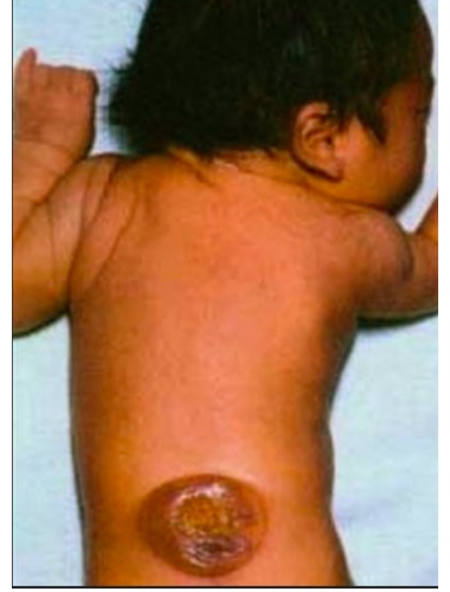
•Meningocele (protruded sac, no neural element)
•Myelomeningocele (protruded sac with neural elements)
•Rachischisis (neural elements exposed)
Cause of neurological deterioration:
•- Tethered cord, hydrocephalus, syringomyelia
Presentation:
•Deformity due to imbalanced muscle pull (hip dislocation, ankle deformity)
•Contracture
•Scoliosis (congenital / muscle imbalance / secondary to unilateral hip dislocation with LLD)
•Functional deficit (Walking difficulty)
•Neurogenic bladder
•Foot deformities
•Assess last functional level
•Walk? (yes at least L3)
•Ankle equinus? (no at least L4)
•Hip abduction? (yes at least L5)
•If not walk: check hip control (no L1 or above, yes with flexion L2)
| Type / group | Definition / note |
|---|---|
| L3 | highest risk of hip dislocation (intact adductor but weak abductor), household walking |
Treatment: depends on form, level
Aim:
•Maximize patient’s function consistent with the level involved and prevent deterioration
Multidisciplinary approach
•Patient and family counselling
Community based treatment and minimize hospital admission
•Use of orthosis
•Physiotherapy for stretching to prevent contracture, maintain muscle power
•Surgery: balance muscle pull to prevent development of deformity/ correct deformity
•Hip: highest dislocation rate in L3,4 last functional level due to imbalance of adductor and abductor. Surgery is controversial and only applicable in patient with functional quadriceps
•Knee: flexion contracture release if severe and affecting walking
•Ankle: can be equinovarus, cavovarus, calcaneovalgus