FRCS Paediatric Ortho — improved notes
FRCS Paediatric Ortho — visual inline notes

121. Paediatric radial neck fracture

visual-inline rebuild2 extracted figuresspeaker notes XML32 study lines4 Q&A

Individual slide images have been cropped from the rendered slide and inserted beside the matching note section. The full slide remains below only for layout/audit.

Clean FRCS notes with inline figures

Describe Xray: Fracture radial neck with >30deg angulation
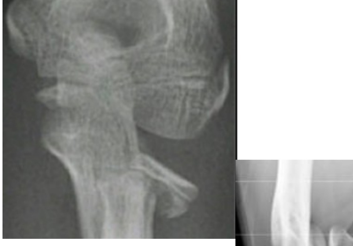
Radiograph / imaging
Fig 1. Radiograph / imagingtop left
Radiograph / imaging
Fig 2. Radiograph / imagingmiddle centre

Obrien classification

•I <30
•II 30-60
•III >60
•Median age is 9-10 years
•Usually associated with a valgus loading injury of the elbow
•Associated Conditions
•Elbow dislocation
•Medial epicondyle fracture

Xray

•Greenspan (radiocapitellar view: elbow 90deg flexion, thumb up, beam 45 from table)

Treatment:

•Depends on angulation
•<30deg: immobilization in situ
•>30deg: attempt close reduction
•Patterson (traction, varus stress at elbow, thumb direct pressure on radial head)
•Israeli (elbow supination + flexion, finger pressure at radial head, pronate to force radial head through)
•Chambers (Esmarch applied distal to proximal, reduction by circumferential pressure

If post reduction still >30deg:

•Percutaneous reduction with K wire (Joystick technique)
•Metaizeau technique: retrograde IM Kwire / TENS with bent tip to lever radial head fragment
•Open reduction has been associated with a greater loss of motion, increased rates of osteonecrosis and synostosis compared with closed reduction

Complication:

•Overgrowth (more common)
•Radial head AVN
•RU synostosis
•Myositis ossificans

•Normal radial neck angulation
•0-15 lateral
•5 post to 10 anterior

Direct Q&A

Q1 How is Paediatric radial neck fracture classified here?
•I <30
•II 30-60
•III >60
•Median age is 9-10 years
•Usually associated with a valgus loading injury of the elbow
•Associated Conditions
•Elbow dislocation
•Medial epicondyle fracture
Q2 What are the radiographic findings for Paediatric radial neck fracture?
•Greenspan (radiocapitellar view: elbow 90deg flexion, thumb up, beam 45 from table)
Q3 How is Paediatric radial neck fracture managed?

Depends on angulation

•<30deg: immobilization in situ
•>30deg: attempt close reduction
•Patterson (traction, varus stress at elbow, thumb direct pressure on radial head)
•Israeli (elbow supination + flexion, finger pressure at radial head, pronate to force radial head through)
•Chambers (Esmarch applied distal to proximal, reduction by circumferential pressure
Q4 What complications are listed for Paediatric radial neck fracture?

Overgrowth (more common)

•Radial head AVN
•RU synostosis
•Myositis ossificans

Raw speaker-note hierarchy audit

#lvlmarLindentlogicaloriginal note line
10NoneNone0Describe Xray: Fracture radial neck with >30deg angulation
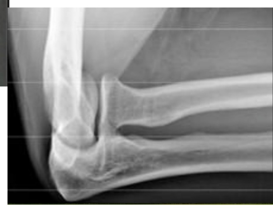
20NoneNone0Obrien classification
30171450-1714501I <30
40171450-1714501II 30-60
50171450-1714501III >60
60NoneNone0Median age is 9-10 years
70NoneNone0Usually associated with a valgus loading injury of the elbow
80NoneNone0Associated Conditions
90171450-1714501Elbow dislocation
100171450-1714501Medial epicondyle fracture
110000Xray
120000Greenspan (radiocapitellar view: elbow 90deg flexion, thumb up, beam 45 from table)
130000Treatment:
140000Depends on angulation
150000<30deg: immobilization in situ
160000>30deg: attempt close reduction
170171450-1714501Patterson (traction, varus stress at elbow, thumb direct pressure on radial head)
180171450-1714501Israeli (elbow supination + flexion, finger pressure at radial head, pronate to force radial head through)
190171450-1714501Chambers (Esmarch applied distal to proximal, reduction by circumferential pressure
200000If post reduction still >30deg:
210171450-1714501Percutaneous reduction with K wire (Joystick technique)
220171450-1714501Metaizeau technique: retrograde IM Kwire / TENS with bent tip to lever radial head fragment
230000Open reduction has been associated with a greater loss of motion, increased rates of osteonecrosis and synostosis compared with closed reduction
240000Complication:
250171450-1714501Overgrowth (more common)
260171450-1714501Radial head AVN
270171450-1714501RU synostosis
280171450-1714501Myositis ossificans
290000==================================
300000Normal radial neck angulation
3100000-15 lateral
3200005 post to 10 anterior

Improved semantic hierarchy used for the study notes

#semantic levelsection?rewritten line
10yesDescribe Xray: Fracture radial neck with >30deg angulation
20yesObrien classification
31I <30
41II 30-60
51III >60
61Median age is 9-10 years
71Usually associated with a valgus loading injury of the elbow
81Associated Conditions
91Elbow dislocation
101Medial epicondyle fracture
110yesXray
121Greenspan (radiocapitellar view: elbow 90deg flexion, thumb up, beam 45 from table)
130yesTreatment:
141Depends on angulation
151<30deg: immobilization in situ
161>30deg: attempt close reduction
171Patterson (traction, varus stress at elbow, thumb direct pressure on radial head)
181Israeli (elbow supination + flexion, finger pressure at radial head, pronate to force radial head through)
191Chambers (Esmarch applied distal to proximal, reduction by circumferential pressure
200yesIf post reduction still >30deg:
211Percutaneous reduction with K wire (Joystick technique)
221Metaizeau technique: retrograde IM Kwire / TENS with bent tip to lever radial head fragment
231Open reduction has been associated with a greater loss of motion, increased rates of osteonecrosis and synostosis compared with closed reduction
240yesComplication:
251Overgrowth (more common)
261Radial head AVN
271RU synostosis
281Myositis ossificans
290——
301Normal radial neck angulation
3110-15 lateral
3215 post to 10 anterior