FRCS Paediatric Ortho — improved notes
FRCS Paediatric Ortho — visual inline notes

122. Paediatric lateral condyle fracture

visual-inline rebuild1 extracted figuresspeaker notes XML53 study lines7 Q&A

Individual slide images have been cropped from the rendered slide and inserted beside the matching note section. The full slide remains below only for layout/audit.

Clean FRCS notes with inline figures

•Stem: Patient with fall injury + axial loading to UL, lateral side of elbow pain + swelling

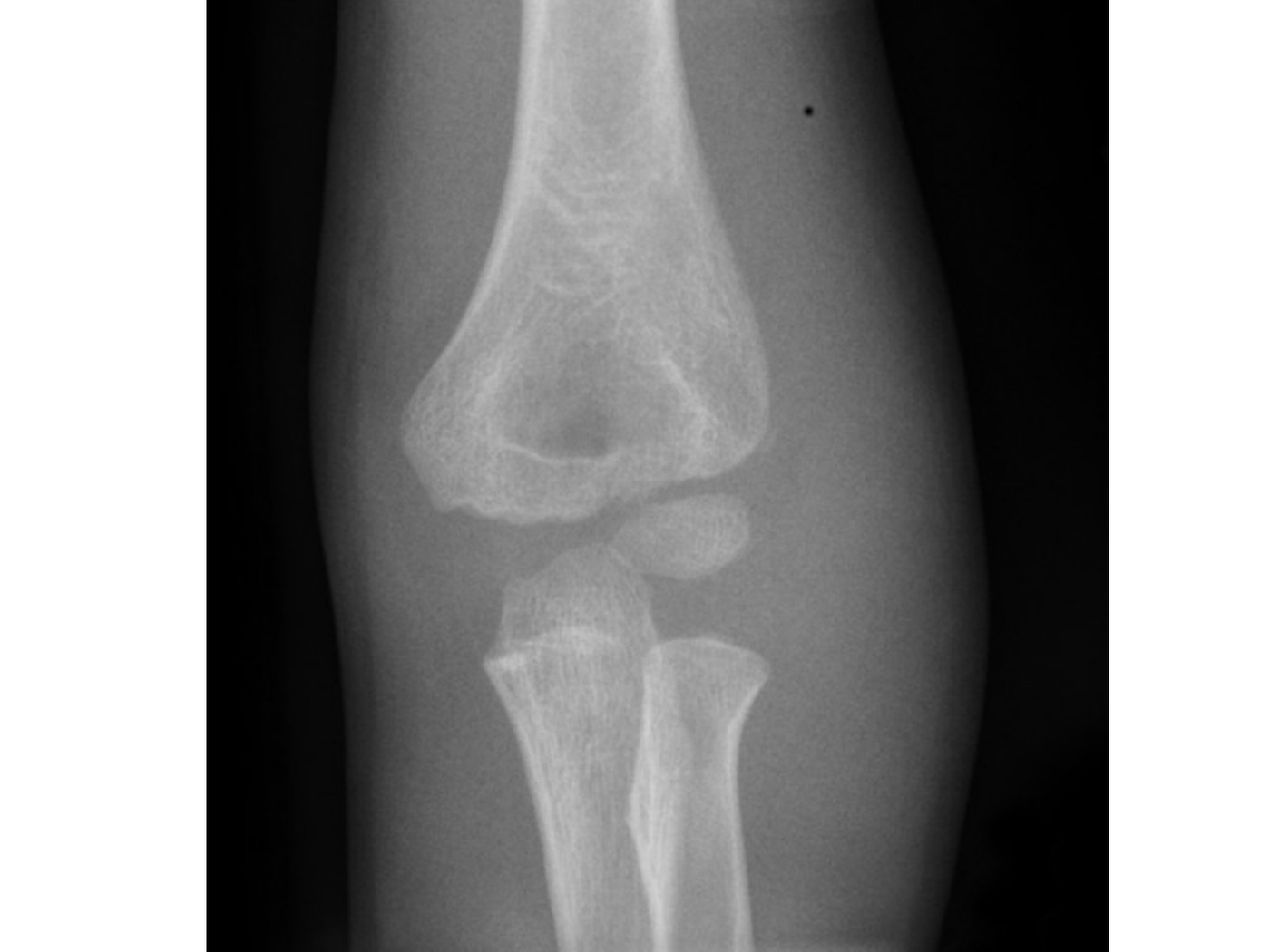
Radiographic description

•Flake of bone over lateral metaphyseal region, no gross mal alignment
•Suspected lateral condyle fracture
•This fracture on Xray is minimally displaced. But I will need to see the articular surface displacement before planning Mx. Can also get an internal oblique XR
•In my centre MRI is readily available, will book urgent MRI to look for articular step.
Arthrogram image
Fig 1. Arthrogram imagetop left
•If MRI is not available, I can perform arthrogram (inject from posterior to avoid injury to articular surface and major NV bundle)

Classification

Milch classification (stability)

Type / groupDefinition / note
1fracture lateral to trochleocapitellar groove, SH type 4, more stable
2fracture medial to trochleocapitellar groove, SH type 2 (no cross lateral condyle epiphysis), less stable → worse prognosis
•Jacob (Displacement)
•1 <2mm displacement (can go for conservative if confirmed articular cartilage smooth)
•2 2-4mm displacement (CR +/-
•OR KWF)
•3 severe displacement +/- rotated fragment (OR KWF)
•Another recently validated classification is the Song classification (1-5), 3 or above unstable, need surgery
•1 – limited fracture line within the metaphysis
•2 – lateral gap
•3 - medial and lateral gap
•4 – more than 2mm displacement with no rotation
•5 – rotation

Definitive management

•If there is >2mm displacement in any of the 3 views, will go for OR + KWF
•CR method is by extension + supination to relax muscle pull from common extensor origin. Then varus force + anteromedial force to fracture to reduce it.
•OR: oblique wound over lateral side centered at lateral epicondyle (between BR and triceps),
•[OB: below the skin, dissection to the joint is most often already accomplished by injury]
•avoid dissection to posterior muscle to preserve blood supply.
•Check anterior and lateral cortex smoothness to confirm reduction (not ossified on XR)
•Fix with 2 divergent pins (reduce with joystick method)

Late complication:

•Nonunion, AVN
•Deformity
•Varus due to lateral condyle overgrowth or fish tail deformity (OB: up to 50%)
•Valgus due to lateral condyle AVN and nonunion (OB: 10%)
•Instability (PL subluxation of proximal radius and ulnar)
•If late presentation and cannot reduce without stripping posterior periosteum, may need to accept partially reduced alignment

Mechanism:

•Push off (axial loading by radial head)
•Pull off (common extensor)

Why prone to nonunion?

•poor vascular supply, intra-articular, large pull from extensor

Delay presentation with nonunion with deformity, no pain. What is your management? Will you perform OT?

•Controversial
•Yes due to future valgus + instability + tardy ulnar nerve palsy (Masada JBJS 1990)
•Preop CT to assess fragment size, MRI to rule out AVN

Aim: achieve union

•Debridement + bone graft + screw horizontally (K wire cannot give compression)
•Only debride anterior, leave posterior intact
•Don’t aggressively reduce, risk of AVN and fishtail deformity
•No, can have osteotomy later, watchful waiting. ORIF associated with stiffness, AVN (Jakob et al)

Recent studies suggest surgery is a safe option

Direct Q&A

Q1 What are the radiographic findings for Paediatric lateral condyle fracture?
•Flake of bone over lateral metaphyseal region, no gross mal alignment
•Suspected lateral condyle fracture
•This fracture on Xray is minimally displaced. But I will need to see the articular surface displacement before planning Mx. Can also get an internal oblique XR
•In my centre MRI is readily available, will book urgent MRI to look for articular step.
•If MRI is not available, I can perform arthrogram (inject from posterior to avoid injury to articular surface and major NV bundle)
Q2 How is Paediatric lateral condyle fracture classified here?
•1: fracture lateral to trochleocapitellar groove, SH type 4, more stable
•2: fracture medial to trochleocapitellar groove, SH type 2 (no cross lateral condyle epiphysis), less stable → worse prognosis
•Jacob (Displacement)
•1 <2mm displacement (can go for conservative if confirmed articular cartilage smooth)
•2 2-4mm displacement (CR +/-
•OR KWF)
•3 severe displacement +/- rotated fragment (OR KWF)
•Another recently validated classification is the Song classification (1-5), 3 or above unstable, need surgery
•1 – limited fracture line within the metaphysis
•2 – lateral gap
•3 - medial and lateral gap
•4 – more than 2mm displacement with no rotation
•5 – rotation
Q3 What is the definitive management for Paediatric lateral condyle fracture?
•If there is >2mm displacement in any of the 3 views, will go for OR + KWF
•CR method is by extension + supination to relax muscle pull from common extensor origin. Then varus force + anteromedial force to fracture to reduce it.
•OR: oblique wound over lateral side centered at lateral epicondyle (between BR and triceps),
•[OB: below the skin, dissection to the joint is most often already accomplished by injury]

avoid dissection to posterior muscle to preserve blood supply.

•Check anterior and lateral cortex smoothness to confirm reduction (not ossified on XR)
•Fix with 2 divergent pins (reduce with joystick method)
Q4 What complications are listed for Paediatric lateral condyle fracture?

Nonunion, AVN

•Deformity
•Varus due to lateral condyle overgrowth or fish tail deformity (OB: up to 50%)
•Valgus due to lateral condyle AVN and nonunion (OB: 10%)
•Instability (PL subluxation of proximal radius and ulnar)
•If late presentation and cannot reduce without stripping posterior periosteum, may need to accept partially reduced alignment
Q5 Why prone to nonunion?

poor vascular supply, intra-articular, large pull from extensor

Q6 Delay presentation with nonunion with deformity, no pain. What is your management? Will you perform OT?

Controversial

•Yes due to future valgus + instability + tardy ulnar nerve palsy (Masada JBJS 1990)
•Preop CT to assess fragment size, MRI to rule out AVN

Aim: achieve union

•Debridement + bone graft + screw horizontally (K wire cannot give compression)
•Only debride anterior, leave posterior intact
•Don’t aggressively reduce, risk of AVN and fishtail deformity
•No, can have osteotomy later, watchful waiting. ORIF associated with stiffness, AVN (Jakob et al)
Q7 What are the management aims for Paediatric lateral condyle fracture?
•Debridement + bone graft + screw horizontally (K wire cannot give compression)
•Only debride anterior, leave posterior intact
•Don’t aggressively reduce, risk of AVN and fishtail deformity
•No, can have osteotomy later, watchful waiting. ORIF associated with stiffness, AVN (Jakob et al)

Raw speaker-note hierarchy audit

#lvlmarLindentlogicaloriginal note line
10000Stem: Patient with fall injury + axial loading to UL, lateral side of elbow pain + swelling
20NoneNone0Describe Xray:
30NoneNone0Flake of bone over lateral metaphyseal region, no gross mal alignment
40NoneNone0Suspected lateral condyle fracture
50000This fracture on Xray is minimally displaced. But I will need to see the articular surface displacement before planning Mx. Can also get an internal oblique XR
60000In my centre MRI is readily available, will book urgent MRI to look for articular step.
70000If MRI is not available, I can perform arthrogram (inject from posterior to avoid injury to articular surface and major NV bundle)
80000Classification
90000Milch classification (stability)
100171450-17145011: fracture lateral to trochleocapitellar groove, SH type 4, more stable
110171450-17145012: fracture medial to trochleocapitellar groove, SH type 2 (no cross lateral condyle epiphysis), less stable → worse prognosis
120000Jacob (Displacement)
130171450-17145011 <2mm displacement (can go for conservative if confirmed articular cartilage smooth)
140171450-17145012 2-4mm displacement (CR +/-
150171450-1714501OR KWF)
160171450-17145013 severe displacement +/- rotated fragment (OR KWF)
170000Another recently validated classification is the Song classification (1-5), 3 or above unstable, need surgery
1800001 – limited fracture line within the metaphysis
1900002 – lateral gap
2000003 - medial and lateral gap
2100004 – more than 2mm displacement with no rotation
2200005 – rotation
230000Definitive management
240000If there is >2mm displacement in any of the 3 views, will go for OR + KWF
250000CR method is by extension + supination to relax muscle pull from common extensor origin. Then varus force + anteromedial force to fracture to reduce it.
260000OR: oblique wound over lateral side centered at lateral epicondyle (between BR and triceps),
270000[OB: below the skin, dissection to the joint is most often already accomplished by injury]
280000avoid dissection to posterior muscle to preserve blood supply.
290000Check anterior and lateral cortex smoothness to confirm reduction (not ossified on XR)
300000Fix with 2 divergent pins (reduce with joystick method)
310NoneNone0Late complication:
320NoneNone0Nonunion, AVN
330NoneNone0Deformity
340000Varus due to lateral condyle overgrowth or fish tail deformity (OB: up to 50%)
351NoneNone1Valgus due to lateral condyle AVN and nonunion (OB: 10%)
360NoneNone0Instability (PL subluxation of proximal radius and ulnar)
370NoneNone0If late presentation and cannot reduce without stripping posterior periosteum, may need to accept partially reduced alignment
380000================================
390NoneNone0Mechanism:
400171450-1714501Push off (axial loading by radial head)
410171450-1714501Pull off (common extensor)
420000Why prone to nonunion?
430000poor vascular supply, intra-articular, large pull from extensor
440000Delay presentation with nonunion with deformity, no pain. What is your management? Will you perform OT?
450171450-1714501Controversial
460171450-1714501Yes due to future valgus + instability + tardy ulnar nerve palsy (Masada JBJS 1990)
471628650-1714502Preop CT to assess fragment size, MRI to rule out AVN
481628650-1714502Aim: achieve union
491628650-1714502Debridement + bone graft + screw horizontally (K wire cannot give compression)
5021085850-1714503Only debride anterior, leave posterior intact
511628650-1714502Don’t aggressively reduce, risk of AVN and fishtail deformity
520171450-1714501No, can have osteotomy later, watchful waiting. ORIF associated with stiffness, AVN (Jakob et al)
530171450-1714501Recent studies suggest surgery is a safe option

Improved semantic hierarchy used for the study notes

#semantic levelsection?rewritten line
10Stem: Patient with fall injury + axial loading to UL, lateral side of elbow pain + swelling
20yesRadiographic description
31Flake of bone over lateral metaphyseal region, no gross mal alignment
41Suspected lateral condyle fracture
51This fracture on Xray is minimally displaced. But I will need to see the articular surface displacement before planning Mx. Can also get an internal oblique XR
61In my centre MRI is readily available, will book urgent MRI to look for articular step.
71If MRI is not available, I can perform arthrogram (inject from posterior to avoid injury to articular surface and major NV bundle)
80yesClassification
90yesMilch classification (stability)
1011: fracture lateral to trochleocapitellar groove, SH type 4, more stable
1112: fracture medial to trochleocapitellar groove, SH type 2 (no cross lateral condyle epiphysis), less stable → worse prognosis
121Jacob (Displacement)
1311 <2mm displacement (can go for conservative if confirmed articular cartilage smooth)
1412 2-4mm displacement (CR +/-
151OR KWF)
1613 severe displacement +/- rotated fragment (OR KWF)
171Another recently validated classification is the Song classification (1-5), 3 or above unstable, need surgery
1811 – limited fracture line within the metaphysis
1912 – lateral gap
2013 - medial and lateral gap
2114 – more than 2mm displacement with no rotation
2215 – rotation
230yesDefinitive management
241If there is >2mm displacement in any of the 3 views, will go for OR + KWF
251CR method is by extension + supination to relax muscle pull from common extensor origin. Then varus force + anteromedial force to fracture to reduce it.
261OR: oblique wound over lateral side centered at lateral epicondyle (between BR and triceps),
271[OB: below the skin, dissection to the joint is most often already accomplished by injury]
281avoid dissection to posterior muscle to preserve blood supply.
291Check anterior and lateral cortex smoothness to confirm reduction (not ossified on XR)
301Fix with 2 divergent pins (reduce with joystick method)
310yesLate complication:
321Nonunion, AVN
331Deformity
341Varus due to lateral condyle overgrowth or fish tail deformity (OB: up to 50%)
351Valgus due to lateral condyle AVN and nonunion (OB: 10%)
361Instability (PL subluxation of proximal radius and ulnar)
371If late presentation and cannot reduce without stripping posterior periosteum, may need to accept partially reduced alignment
380——
390yesMechanism:
401Push off (axial loading by radial head)
411Pull off (common extensor)
420yesWhy prone to nonunion?
431poor vascular supply, intra-articular, large pull from extensor
440yesDelay presentation with nonunion with deformity, no pain. What is your management? Will you perform OT?
451Controversial
461Yes due to future valgus + instability + tardy ulnar nerve palsy (Masada JBJS 1990)
472Preop CT to assess fragment size, MRI to rule out AVN
482yesAim: achieve union
493Debridement + bone graft + screw horizontally (K wire cannot give compression)
503Only debride anterior, leave posterior intact
512Don’t aggressively reduce, risk of AVN and fishtail deformity
521No, can have osteotomy later, watchful waiting. ORIF associated with stiffness, AVN (Jakob et al)
530yesRecent studies suggest surgery is a safe option