FRCS Paediatric Ortho — improved notes
FRCS Paediatric Ortho — visual inline notes

123. Post lateral condyle fracture deformity

visual-inline rebuild2 extracted figuresspeaker notes XML36 study lines3 Q&A

Individual slide images have been cropped from the rendered slide and inserted beside the matching note section. The full slide remains below only for layout/audit.

Clean FRCS notes with inline figures

Blood supply of distal humerus

•Radial recurrent artery (comes from radial collateral artery)
•Interosseous recurrent artery (comes from middle collateral artery)
•Lateral condyle epiphysis
•Entry: posterolateral to origin of capsule and proximal to articular cartilage (end artery)
•Trochlea
•Lateral crista: from lateral condyle

Medial crista:

•Lateral part: across posterior surface of distal humerus metaphysis (end artery)
•Medial part: multiple vessels
•Fishtail deformity
•Area between medial ossification center and lateral condyle ossification center resorbs / fails to develop
•Type 1
•More common
•Sharp angled wedge
•Persistent gap between lateral condyle physis and medial trochlear due to underdevelopment of the lateral crista
•Type 2
•Smooth edge
•AVN of lateral part of medial crista of trochlear
•Varus
•1. Overgrowth (50%)
•2. Fishtail
•Malunion of lateral part of medial crista (type 1)
•Medial crista AVN (type 2)
•Valgus
•1. Nonunion → lateral condyle AVN
•2. Physeal arrest
•Nonunion
•Definition: 3 months
•Displaced >2mm + no K wire fixation → lateral condyle AVN

Management

•Before physeal closure → Osteotomy + BG (displaced <1cm, large fragment)
•After physeal closure
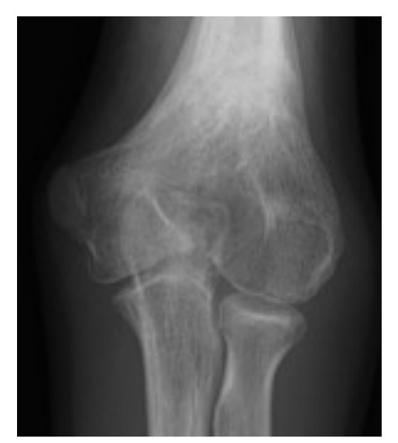
Ultrasound image
Fig 1. Ultrasound imagemiddle left
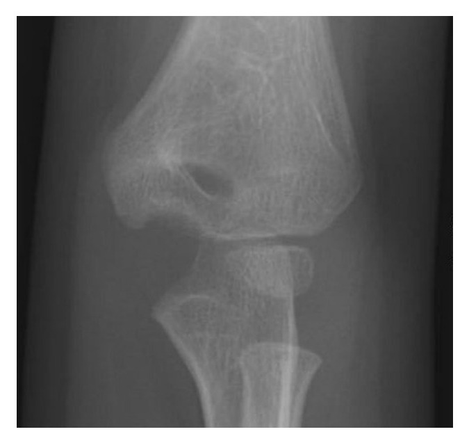
Ultrasound image
Fig 2. Ultrasound imagemiddle centre
•Freshen edges + bone graft + compression for fixation
•Severe valgus deformity → osteotomy
•Ulnar nerve palsy → transposition

Direct Q&A

Q1 What diagnostic or radiographic findings support Post lateral condyle fracture deformity?

Blood supply of distal humerus

•Radial recurrent artery (comes from radial collateral artery)
•Interosseous recurrent artery (comes from middle collateral artery)
•Lateral condyle epiphysis
•Entry: posterolateral to origin of capsule and proximal to articular cartilage (end artery)
•Trochlea
•Lateral crista: from lateral condyle

Medial crista:

•Lateral part: across posterior surface of distal humerus metaphysis (end artery)
•Medial part: multiple vessels
Q2 What blood supply pattern is described for Post lateral condyle fracture deformity?

Radial recurrent artery (comes from radial collateral artery)

•Interosseous recurrent artery (comes from middle collateral artery)
•Lateral condyle epiphysis
•Entry: posterolateral to origin of capsule and proximal to articular cartilage (end artery)
•Trochlea
•Lateral crista: from lateral condyle
Q3 How is Post lateral condyle fracture deformity managed?
•Before physeal closure → Osteotomy + BG (displaced <1cm, large fragment)
•After physeal closure
•Freshen edges + bone graft + compression for fixation
•Severe valgus deformity → osteotomy
•Ulnar nerve palsy → transposition

Raw speaker-note hierarchy audit

#lvlmarLindentlogicaloriginal note line
10NoneNone0Blood supply of distal humerus
20171450-1714501Radial recurrent artery (comes from radial collateral artery)
30171450-1714501Interosseous recurrent artery (comes from middle collateral artery)
40NoneNone0Lateral condyle epiphysis
50NoneNone0Entry: posterolateral to origin of capsule and proximal to articular cartilage (end artery)
60NoneNone0Trochlea
70NoneNone0Lateral crista: from lateral condyle
80NoneNone0Medial crista:
91628650-1714502Lateral part: across posterior surface of distal humerus metaphysis (end artery)
101628650-1714502Medial part: multiple vessels
110NoneNone0Fishtail deformity
120NoneNone0Area between medial ossification center and lateral condyle ossification center resorbs / fails to develop
130NoneNone0Type 1
140NoneNone0More common
150NoneNone0Sharp angled wedge
160NoneNone0Persistent gap between lateral condyle physis and medial trochlear due to underdevelopment of the lateral crista
170NoneNone0Type 2
180NoneNone0Smooth edge
190NoneNone0AVN of lateral part of medial crista of trochlear
200NoneNone0Varus
210NoneNone01. Overgrowth (50%)
220NoneNone02. Fishtail
230171450-1714501Malunion of lateral part of medial crista (type 1)
240171450-1714501Medial crista AVN (type 2)
250000Valgus
2600001. Nonunion → lateral condyle AVN
2700002. Physeal arrest
280000Nonunion
290000Definition: 3 months
300000Displaced >2mm + no K wire fixation → lateral condyle AVN
310000Mx:
320000Before physeal closure → Osteotomy + BG (displaced <1cm, large fragment)
330000After physeal closure
340228600-2286001Freshen edges + bone graft + compression for fixation
350228600-2286001Severe valgus deformity → osteotomy
360228600-2286001Ulnar nerve palsy → transposition

Improved semantic hierarchy used for the study notes

#semantic levelsection?rewritten line
10yesBlood supply of distal humerus
21Radial recurrent artery (comes from radial collateral artery)
31Interosseous recurrent artery (comes from middle collateral artery)
41Lateral condyle epiphysis
51Entry: posterolateral to origin of capsule and proximal to articular cartilage (end artery)
61Trochlea
71Lateral crista: from lateral condyle
80yesMedial crista:
92Lateral part: across posterior surface of distal humerus metaphysis (end artery)
102Medial part: multiple vessels
111Fishtail deformity
121Area between medial ossification center and lateral condyle ossification center resorbs / fails to develop
131Type 1
141More common
151Sharp angled wedge
161Persistent gap between lateral condyle physis and medial trochlear due to underdevelopment of the lateral crista
171Type 2
181Smooth edge
191AVN of lateral part of medial crista of trochlear
201Varus
2111. Overgrowth (50%)
2212. Fishtail
231Malunion of lateral part of medial crista (type 1)
241Medial crista AVN (type 2)
251Valgus
2611. Nonunion → lateral condyle AVN
2712. Physeal arrest
281Nonunion
291Definition: 3 months
301Displaced >2mm + no K wire fixation → lateral condyle AVN
310yesManagement
321Before physeal closure → Osteotomy + BG (displaced <1cm, large fragment)
331After physeal closure
341Freshen edges + bone graft + compression for fixation
351Severe valgus deformity → osteotomy
361Ulnar nerve palsy → transposition