Clean FRCS notes with inline figures
Monteggia fracture dislocation
•Proximal 1/3 ulnar fracture with associated radial head dislocation / instability
Bado classification
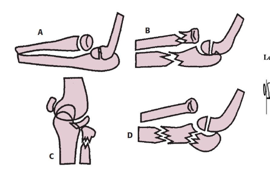
| Type / group | Definition / note |
|---|---|
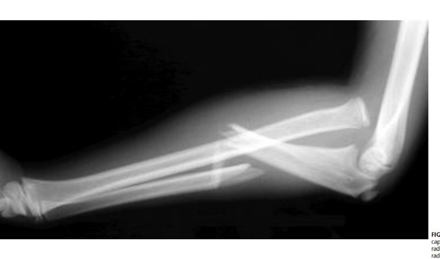
| I | Anterior dislocation + mid-prox 1/3 ulna #, apex anterior. |
•Mechanism: Hyperpronation or hyperextension
•Reduce with flexion
| Type / group | Definition / note |
|---|---|
| II | Posterior dislocation + mid-prox 1/3 ulna #, apex posterior. |
•Mechanism: Axial load of forearm with flexed elbow
•Reduce with extension
| Type / group | Definition / note |
|---|---|
| III | Lateral dislocation + ulnar metaphyseal #. |
•Mechanism: Extension + varus stress
| Type / group | Definition / note |
|---|---|
| IV | Fracture both radius and ulna, anterior dislocation. Type 1 + radial shaft additional fail |
•(children I, adult II)
•Type 1 equivalent (“Monteggia variant”)
| Type / group | Definition / note |
|---|---|
| Type I | isolated radial head dislocation |
| Type II | radial neck fracture + ulna fracture |
| Type III | isolated radial neck fracture |
| Type IV | elbow dislocation |
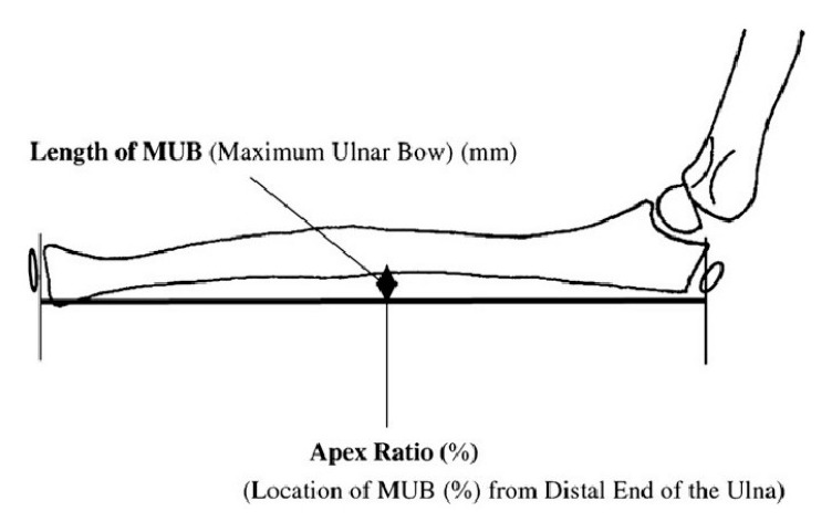
•Ulnar bow line – plastic deformity of ulna
•Ulna should be very straight!
•Line joining proximal and distal ulna on lateral XR, increase ulnar bow if >1cm from apex
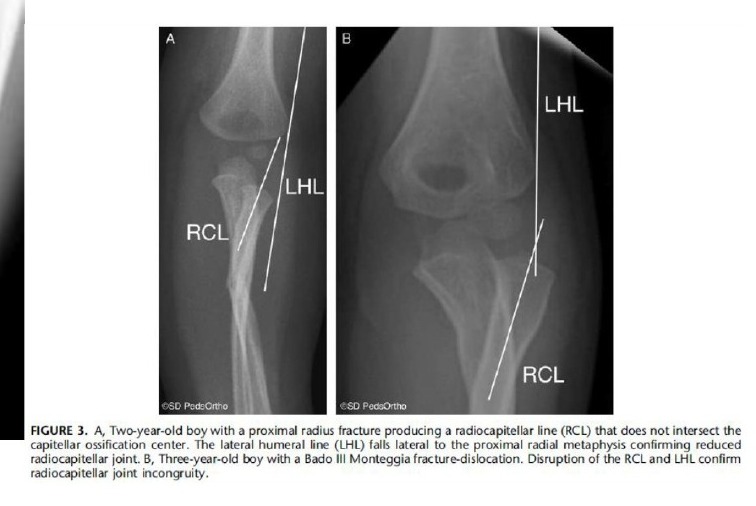
•Lateral humeral line
•Line along axis of humerus, shift to lateral epicondyle, normally it lines lateral to proximal radial metaphysis (medial to it in bado 3)
Acute Management: assess other injury, neurovascular, open wound
•Reduction of ulna fracture (flexion, traction, supination + direct pressure on radial head), maintain length, alignment, radius reduced after ulna reduced + length restored
•Immobilize in supination (tighten IOM) and flexion for type I, extension (<60) in type II
Definitive mx:
•Aim: concentric RCJ, anatomical fixation of ulnar (length, angulation, rotation)
•Fix with plate or K wire + cast (supination in I/ III) for <10yo
•ORIF if >10yo or if fracture is very comminuted or length unstable
•Radial head will usually automatically reduce if ulnar reduction is achieved
•If greenstick fracture of ulna + radial head dislocation
•Try reduce ulna greenstick by sustained force, make use of viscoelastic property of paediatric bone (difficult)
•IM Kwire to fix reduced ulna
•Osteotomy at metaphyseal region to procedure apex dorsal (closer to CORA of radius, not ulna)
•If radial head not reduce
•Confirm ulnar fixation anatomical
•Rule out annular ligament or radial nerve interposition
•Plastic deformity of ulnar
•+/- Bell Tawse for annular ligament reconstruction
+/- Transcapitellar pin (last resort, for subacute case)
•Cx
•Nerve injury
•Most common Bado Type II & III, radial or median nerve, PIN (10%), AIN, observe 3m, if no recovery → explore
•Radial head instability
•< 6 wk + non anatomical ulna reduction → revise fixation + radial head OR
•> 6 wk → osteotomy
Management of late presentation
Sequelae:
•Cubital valgus
•Valgus/ PLRI instability
•Ulnar nerve and PIN palsy
•Heterotopic ossification
•OA
OT indication:
•All <12yo
•Normal concave radial head + convex capitellum
•Ulna osteotomy + open reduction of radial head +/- annular ligament reconstruction
•Cut at ulnar metaphyseal area, closer to site where radius is out; overcorrect due to plastic deformity
Kocher approach for radial head management (protect PIN)
•Check stability
•- Reconstruct annular ligament Bell tawse (lateral strip of triceps fascia wind around radial neck then back onto itself)
•- +/- Transcapitellar Kwire
•- Cast 6w
Complication:
•Residual radial head dislocation
•Ulnar nerve palsy
•Heterotopic ossification
•Radioulnar synostosis
•Nakamura JBJS 2009 showed good long term outcomes after acute recon for chronic missed monteggia fracture under 12yo, within 2 years of initial injury
•JBJS review paper Hubbard 2018
•Timing: duration of dislocation <3yr, patient age <12yo
•Osteotomy: crescentic osteotomy without ulnar lengthening
•Radial head reduction: into native annular ligament, if failed, piecrust, if still fail, incise and repair
•Reinforcement with triceps fascia
•0.4% of all fracture, peak incidence 4-10 yr
Delayed treatment > 2w with much increased complication
•Stabilizers of radial head
Static:
•ligament: IOL (proximal oblique cord) + annular ligament + quadrate ligament
•Bone: RCJ
•Dynamic: Anconeus