FRCS Paediatric Ortho — improved notes
FRCS Paediatric Ortho — visual inline notes

127. Paed forearm fracture

visual-inline rebuild1 extracted figuresspeaker notes XML45 study lines4 Q&A

Individual slide images have been cropped from the rendered slide and inserted beside the matching note section. The full slide remains below only for layout/audit.

Clean FRCS notes with inline figures

Paed forearm fracture
Clinical photograph
Fig 1. Clinical photographtop left
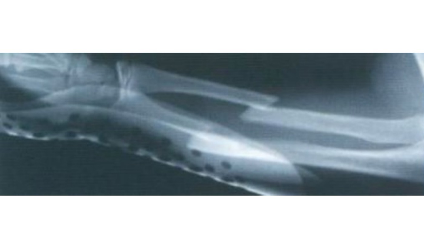
•Describe Xray: fracture of both radius and ulnar at shaft with shortening (bayonet) in skeletally immature patient
•Hx: age, mechanism of injury

r/o acute complication: compartment/ NV injury

Management

•Aim of treatment: restore full rotational range, promote union, avoid complication (malunion, synostosis, refracture)

Factors to consider:

•1. Site of deformity (affect position of cast due to different deforming force)
•2. Degree of deformity
•3. Age of patient (affect deformity we can accept)
•*Acceptable alignment: (Rockwood 2010)
•< 10y.o.: angulation <15, shortening and bayonetting <1cm
•>10y.o.: need anatomical reduction
•<10deg sagittal for 11-13, 0-5deg for >13
•0deg coronal for >=11
•NO rotational malalignment accepted
•Close reduction
•Both bone
•Temp reduce by pointing palm in the direction of apex of deformity
•Traction + angulation + rotation in the direction of angulation
•Bayonette deformity requires more traction to reduce
•Single bone - may need more traction
•Fixation
•K wire fixation – choose appropriately sized k wire (occupy at least 2/3 of diameter of medullary isthmus)
•Prebend radial wire to three times the diameter of the medullary canal and ensure the apex of bend corresponds to level of fracture
•Radial styloid – between EC I and II
•Ulnar olecranon
•Confirm correct rotation

Indication for plating:

•Close to skeletal maturity
•Meta-diaphyseal junction (width mismatch → hard to control by wire)
•Casting method: mold with 3 point fixation to correct angular deformity + AP compression (ligamentotaxis by interosseous membrane), sagittal width: coronal width of cast (Cast index): <0.75 (0.7)

•Deformity force: supinator, pronator teres, pronator quadratus (PNS)
•Proximal 1/3 fracture [prox fragment flexed (biceps) + supinated (supinator), distal pronated (PT +PQ)→ align distal with proximal by supinating)
•Middle 1/3 (proximal balanced by supinator and PT, distal pronated by PQ → align with distal in neutral position)
•Distal 1/3 (proximal pronated by PT + PQ, align with distal in pronation)

How to assess rotation?

•AP: radial styloid/ radial tuberosity 180deg
•Lat: coronoid process/ ulnar styloid 180deg

Complication

•Immediate – compartment syndrome 1-8%
•Malunion (10 deg angulation in 1 bone = 20-30 degrees of rotational block)
•Refracture (5% in 6 months)
•Synostosis

Direct Q&A

Q1 How is Paed forearm fracture managed?
•Aim of treatment: restore full rotational range, promote union, avoid complication (malunion, synostosis, refracture)
Q2 What are the indications listed for Paed forearm fracture?

Close to skeletal maturity

•Meta-diaphyseal junction (width mismatch → hard to control by wire)
•Casting method: mold with 3 point fixation to correct angular deformity + AP compression (ligamentotaxis by interosseous membrane), sagittal width: coronal width of cast (Cast index): <0.75 (0.7)
Q3 How to assess rotation?
•AP: radial styloid/ radial tuberosity 180deg
•Lat: coronoid process/ ulnar styloid 180deg
Q4 What complications are listed for Paed forearm fracture?
•Immediate – compartment syndrome 1-8%
•Malunion (10 deg angulation in 1 bone = 20-30 degrees of rotational block)
•Refracture (5% in 6 months)
•Synostosis

Raw speaker-note hierarchy audit

#lvlmarLindentlogicaloriginal note line
10NoneNone0Paed forearm fracture
20NoneNone0Describe Xray: fracture of both radius and ulnar at shaft with shortening (bayonet) in skeletally immature patient
30NoneNone0Hx: age, mechanism of injury
40000r/o acute complication: compartment/ NV injury
50NoneNone0Mx:
60NoneNone0Aim of treatment: restore full rotational range, promote union, avoid complication (malunion, synostosis, refracture)
70NoneNone0Factors to consider:
80NoneNone01. Site of deformity (affect position of cast due to different deforming force)
90NoneNone02. Degree of deformity
100NoneNone03. Age of patient (affect deformity we can accept)
110NoneNone0*Acceptable alignment: (Rockwood 2010)
120171450-1714501< 10y.o.: angulation <15, shortening and bayonetting <1cm
130171450-1714501>10y.o.: need anatomical reduction
141628650-1714502<10deg sagittal for 11-13, 0-5deg for >13
151628650-17145020deg coronal for >=11
160171450-1714501NO rotational malalignment accepted
170000Close reduction
180000Both bone
190171450-1714501Temp reduce by pointing palm in the direction of apex of deformity
200171450-1714501Traction + angulation + rotation in the direction of angulation
210171450-1714501Bayonette deformity requires more traction to reduce
220000Single bone - may need more traction
230000Fixation
240171450-1714501K wire fixation – choose appropriately sized k wire (occupy at least 2/3 of diameter of medullary isthmus)
250171450-1714501Prebend radial wire to three times the diameter of the medullary canal and ensure the apex of bend corresponds to level of fracture
260171450-1714501Radial styloid – between EC I and II
270171450-1714501Ulnar olecranon
280171450-1714501Confirm correct rotation
290000Indication for plating:
300228600-2286001Close to skeletal maturity
310228600-2286001Meta-diaphyseal junction (width mismatch → hard to control by wire)
320000Casting method: mold with 3 point fixation to correct angular deformity + AP compression (ligamentotaxis by interosseous membrane), sagittal width: coronal width of cast (Cast index): <0.75 (0.7)
330000================
340NoneNone0Deformity force: supinator, pronator teres, pronator quadratus (PNS)
350171450-1714501Proximal 1/3 fracture [prox fragment flexed (biceps) + supinated (supinator), distal pronated (PT +PQ)→ align distal with proximal by supinating)
360171450-1714501Middle 1/3 (proximal balanced by supinator and PT, distal pronated by PQ → align with distal in neutral position)
370171450-1714501Distal 1/3 (proximal pronated by PT + PQ, align with distal in pronation)
380NoneNone0How to assess rotation?
390171450-1714501AP: radial styloid/ radial tuberosity 180deg
400171450-1714501Lat: coronoid process/ ulnar styloid 180deg
410000Complication
420171450-1714501Immediate – compartment syndrome 1-8%
430171450-1714501Malunion (10 deg angulation in 1 bone = 20-30 degrees of rotational block)
440171450-1714501Refracture (5% in 6 months)
450171450-1714501Synostosis

Improved semantic hierarchy used for the study notes

#semantic levelsection?rewritten line
10Paed forearm fracture
21Describe Xray: fracture of both radius and ulnar at shaft with shortening (bayonet) in skeletally immature patient
31Hx: age, mechanism of injury
40yesr/o acute complication: compartment/ NV injury
50yesManagement
61Aim of treatment: restore full rotational range, promote union, avoid complication (malunion, synostosis, refracture)
70yesFactors to consider:
811. Site of deformity (affect position of cast due to different deforming force)
912. Degree of deformity
1013. Age of patient (affect deformity we can accept)
111*Acceptable alignment: (Rockwood 2010)
121< 10y.o.: angulation <15, shortening and bayonetting <1cm
131>10y.o.: need anatomical reduction
142<10deg sagittal for 11-13, 0-5deg for >13
1520deg coronal for >=11
161NO rotational malalignment accepted
171Close reduction
181Both bone
191Temp reduce by pointing palm in the direction of apex of deformity
201Traction + angulation + rotation in the direction of angulation
211Bayonette deformity requires more traction to reduce
221Single bone - may need more traction
231Fixation
241K wire fixation – choose appropriately sized k wire (occupy at least 2/3 of diameter of medullary isthmus)
251Prebend radial wire to three times the diameter of the medullary canal and ensure the apex of bend corresponds to level of fracture
261Radial styloid – between EC I and II
271Ulnar olecranon
281Confirm correct rotation
290yesIndication for plating:
301Close to skeletal maturity
311Meta-diaphyseal junction (width mismatch → hard to control by wire)
321Casting method: mold with 3 point fixation to correct angular deformity + AP compression (ligamentotaxis by interosseous membrane), sagittal width: coronal width of cast (Cast index): <0.75 (0.7)
330——
341Deformity force: supinator, pronator teres, pronator quadratus (PNS)
351Proximal 1/3 fracture [prox fragment flexed (biceps) + supinated (supinator), distal pronated (PT +PQ)→ align distal with proximal by supinating)
361Middle 1/3 (proximal balanced by supinator and PT, distal pronated by PQ → align with distal in neutral position)
371Distal 1/3 (proximal pronated by PT + PQ, align with distal in pronation)
380yesHow to assess rotation?
391AP: radial styloid/ radial tuberosity 180deg
401Lat: coronoid process/ ulnar styloid 180deg
410yesComplication
421Immediate – compartment syndrome 1-8%
431Malunion (10 deg angulation in 1 bone = 20-30 degrees of rotational block)
441Refracture (5% in 6 months)
451Synostosis