Clean FRCS notes with inline figures
•14 y.o boy from china, chronic leg pain after trivial injury
Subacute osteomyelitis
Radiographic description
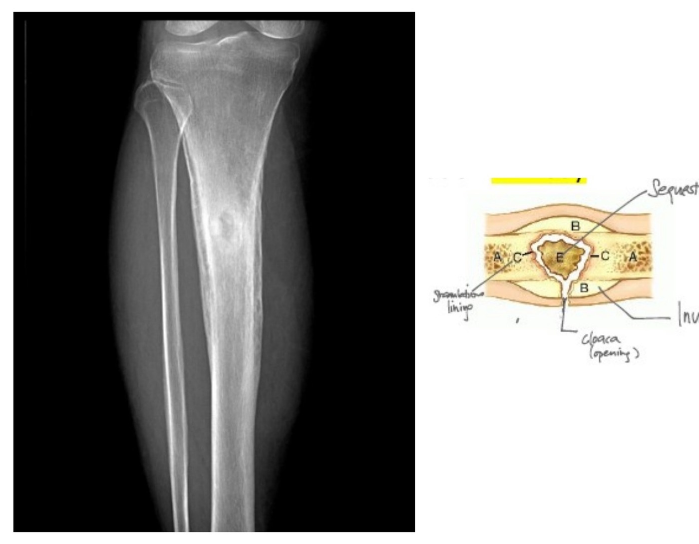
•Lytic lesion: small lytic lesion at proximal diaphysis of leg, concentric and intramedullary, sclerotic rim, narrow transitional zone, significant periosteal reaction
•Ddx: tumour (Ewing in diaphysis, OGS in metaphysis, chondroblastoma in epiphysis), infection (brodies abscess)
•Brodies abscess: chronic abscesses surrounded by sclerotic bone and fibrous tissue
Partially treated acute osteomyelitis, decreased virulence of bacteria
•PE: discharging sinus, skin erythema, LN
•Investigation: blood test (raised ESR 50% +ve, WBC, CRR), MRI, CT
•Cierny staging for chronic osteomyelitis
•Anatomical
•IM
•Superficial
•Local
•Diffuse
•2. Physiological
| Type / group | Definition / note |
|---|---|
| A | healthy |
| B | immunocompromised |
| C | prohibitive |
May need biopsy to confirm diagnosis before OT
•MRI to look for sequestrum
Management principle:
•Similar to chronic OM
•Eradicate infection, reconstruction of bone and soft tissue defects, minimize complications and hospital stay
•Eradication of infection (3)
•1. Debridement
•Saucerization and sequestrectomy, until healthy bleeding (paprika sign)
•Dead space management by antibiotic loaded cement spacer / gentamicin beads / VAC dressing
•2. Antibiotics: local abx loaded cement and systemic antibiotics
•3. +/- skeletal stabilization (depends on Cierny staging)
•Reconstruction
•Bone defect
•Soft tissue
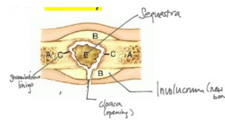
Stages of acute osteomyelitis + draw
•Inflammation
•Suppuration (subperiosteal abscess)
•Necrosis (sequestrum)
•New bone formation (involucum)
•Resolution