Clean FRCS notes with inline figures
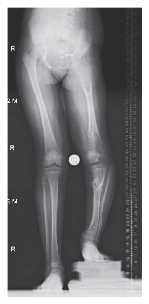
Ollier’s disease
•Sporadic
•Defect enchondral ossification
Clinical:
•Unilateral involvement → LLD
•Angular deformity
•Ddx: fibrous dysplasia (polyostotic), OI
•XR: cartilage containing lesion + irregular translucent area at metaphysis. Expanded bone. Polyostotic
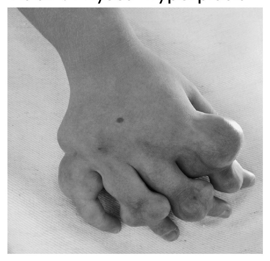
•Maffucci syndrome
•= Ollier + hemangioma
•Expanded bone
•Cutaneous haemangiomata
XR: Ollier + small rounded calcified phleboliths
•Malignant transformation
•Solitary <1%
•Olliers 25-30%
•Maffucci 100%
What to consider in the treatment?
Deformity in 4 planes?
•Coronal: valgus varus
•Sagittal: flexion extension
•Transverse: torsional
•Longitudinal: LLD
•Aim to have well aligned limbs in all those planes
Options:
•Use of hemiepiphysiodesis or guided growth
Osteotomy for acute or gradual correction
•Combo of techniques
•Enchondroma = type of incomplete enchondral ossification, escape of chondroblast to metaphysis (presence of cartilage in IM canal)
•Rubin: physis hyperplasia
•Vs
•Osteochondroma= benign chondrogenic lesion derived from the perichondral ring - metaphysis hyperplasia