Clean FRCS notes with inline figures
MHE
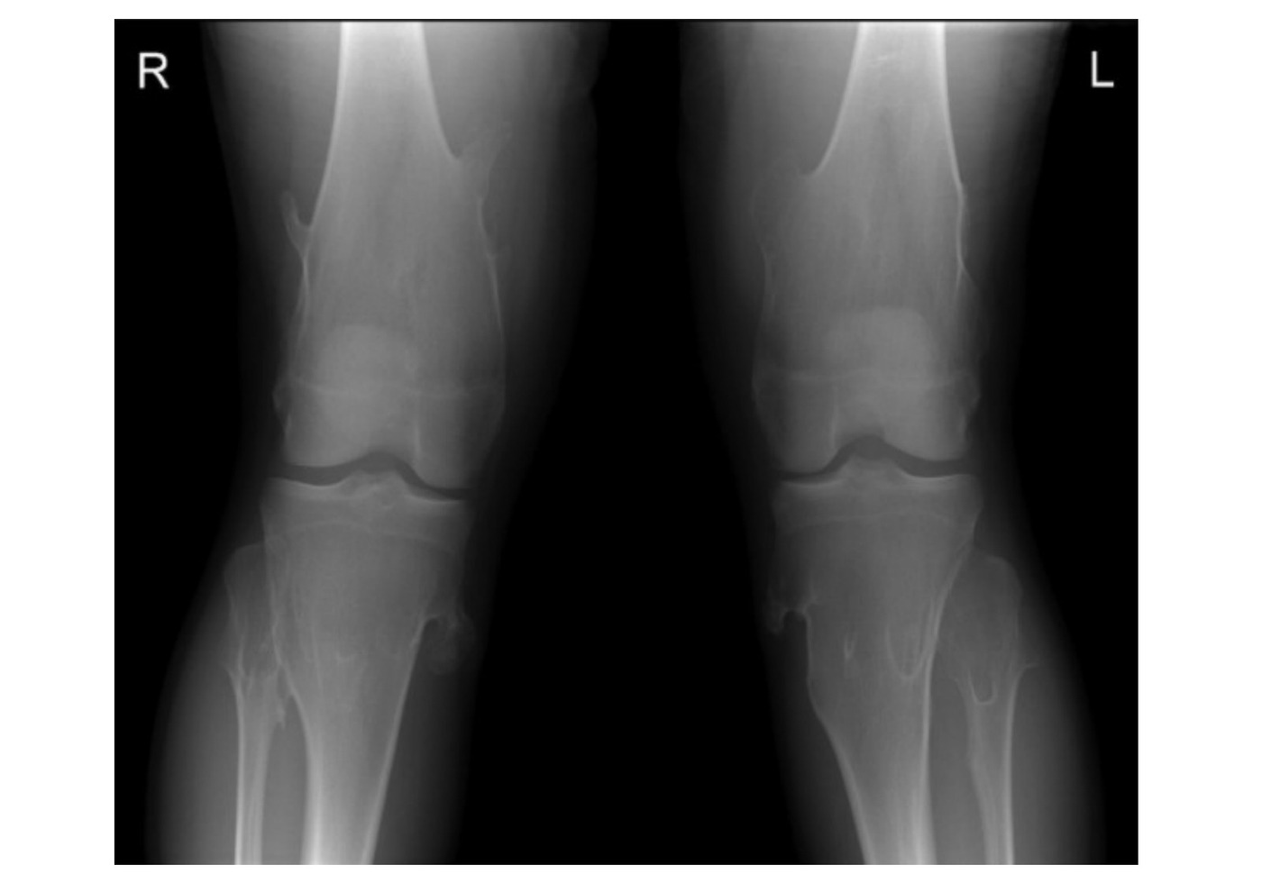
•XR of both knees showing a skeletally mature patient with multiple bony protrusions of the metaphyseal region of distal femur and proximal tibia with associated tubulation of the metaphyseal region.
•The protrusions are pedunculated, well defined, with a narrowing zone of transition and pointing away from the physis. Trabeculae are seen within the lesion and are continuous with the rest of the bone.
•Otherwise there are no cortical destruction, no calcification within the soft tissue indicating a malignant transformation.
•Dx Multiple hereditary exostosis
•Ddx: Trevor disease (dysplasia epiphysealis hemimelia): Epiphyseal exostosis
How to differentiate?
•Clinically: Trevor younger
•Radiological: Trevor arise from epiphysis
•Histological: cartilage cap of Trevor contain bands of cartilage separating areas of cancellous bone
•Gene: normal EXT levels
•Also check clinically the forearms to see if there are any bony exostosis and associated radial head dislocation and bowing
Investigations
•Further XR to look at the scapular and pelvis (regions where clinically cannot be palpated)
•Scanogram for LL alignment
Management
•Benign condition
Rule out complication
•Local pressure sx (nerve, tendon, bursitis, pseudoaneurysm)
•Reduce joint ROM
•Deformity / growth disturbance
•Malignancy
•Stop growing at maturity
•Monitor → 5-10% lifetime risk of malignant transformation (c/w normal lifetime risk of any malignancy of 1 in 3)
Indication for surgery
•Pain from bursitis / affect joint movement / compression on NV and tendon
•Deformity (bowing of forearm, genu varum and valgum), intraarticular exostosis
•Malignant transformation into chondrosarcoma (@ 5th decade) 1% lifetime risk per lesion, thickness >2cm (normal 2-3mm)
•Clinical: Increase in size after puberty, acute onset of pain in adults
•Radiological: cortical destruction, soft tissue mineralization >2cm, MRI cartilage cap >2cm, sessile, central more malignancy
Principle of surgery:
•Excise lesion including cartilaginous cap to reduce recurrence
•Corrective osteotomy +/- lengthening procedures
•MHE is a benign condition with multiple chondrogenic tumor (Defect in the groove of Ranvier, aberrant cartilage from the perichondral ring, mutations affecting prehypertrophic chondrocytes)
•Sessile / pedunculated growth from physeal plate away from joint
•AD, EXT1 exostosin (Chr 8), 2 (Chr 11), 3 gene (Chr 19) (1 is worst, more exostosis, more sarcoma, more limb deformities, more axial bone involved)
•10% spontaneous mutation