FRCS Paediatric Ortho — improved notes
FRCS Paediatric Ortho — visual inline notes

077. Osteogenesis imperfecta OI

visual-inline rebuild1 extracted figuresspeaker notes XML73 study lines5 Q&A

Individual slide images have been cropped from the rendered slide and inserted beside the matching note section. The full slide remains below only for layout/audit.

Clean FRCS notes with inline figures

Osteogenesis imperfecta OI

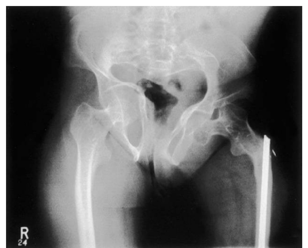
•Xray: Trifoil shaped pelvis, protrusio, lateral bowing of R femur shaft.
•Also check/perform check the rest of the lower limbs, expect to see gracile or overtubulated bones, anterior bowing of tibia (Saber shin), lateral bowing and erlenmeyer flask appearance of femur. In the spine, may find codfish vertebrae or kyphoscoliosis. Also need XR skull to see if there is Wormian bone + basilar invagination.

If fracture:

•Hypertrophic tumoral callus
•Malunion
•Pseudoarthrosis

Diagnosis: OI

•Ddx: rickets/ osteomalacia, idiopathic juvenile osteoporosis, NAI, fibrous dysplasia
•(Ddx for children with repeated fracture)

Examination

•Short stature (proportionate)
•Thin skin
•Face
•Eyes: Blue sclera
•Ears: Impaired hearing (in type 1)
•Mouth: Defective detinogenesis. High pitch voice
•Triangular shaped head with broad forehead

Ortho:

•Lateral bowing of femur
•Anterior bowing of tibia (saber shin)
•Genu varum
•Acetabular protrusion
•Kyphoscoliosis
•Congenital radial head dislocation
•Basilar invagination (myelopathy)
•Generalized ligamentous laxity
•Diagnosis is based on radiological and clinical features +/- family history
•Skin Biopsy is gold standard (culturing dermal fibroblasts)

Treatment Goal

•Prevent fracture
•Deformity correction
•Optimize function
•1. Refer to geneticist to establish diagnosis
•2. Medical:
•Bisphosphonates (decrease fracture: 90% increase in cortical diameter, 50% increase in cancellous bone, decrease pain, increase ambulation)
•Growth hormone
•Ca Vit D
•Gene therapy
•3. Ortho

Fracture:

•Normal healing rate, but no remodeling. Also associated with an increased risk of further fracture due to immobilization and/or the stress riser effect at the edges of the cast
•Therefore, if recurrent fracture → I would treat with CR + IMN +/- multiple osteotomies (Sofield-Miller procedure) to correct deformity at the same time

Deformity correction:

•Limbs: Sofield-Miller with telescoping IM system such as Bailey Dubow rod (if medullary canal is wide enough) / Rush pin
•→ Deformity will progress if left untreated due to the HV law leading to selective inhibition of the physeal growth
•Spine: Scoliosis surgery if cobb >45 (no brace due to rib fragility)
•Sofield-Miller: Do osteotomy first and connect with IMN
•Modified Sofield-Miller: Insert IM rod till resistance and osteotomy at that site

Difficulties:

•Anes – malignant hyperthermia, basilar invagination, mitral valve regurg
Ultrasound image
Fig 1. Ultrasound imagetop left
•Ortho – bleeding, abnormal bone quality, minimal autograft, ligamentous laxity

•Osteogenesis Imperfecta

Rubin classification: Diaphyseal hypoplasia

•Hereditary disease with abnormal Type 1 collagen (quantitative/ qualitative)
•90% have identifiable genetic mutation
•Both quantitative and qualitative defect in COL1A1 and COL1A2 [substitution of glycine by a bulky amino acid e.g. cystine in the procollagen structure, unable to crosslink], leads to reduced collagen secretion and abnormal collagen production → insufficient osteoid production. Unable to remodel normally. But initial fracture healing is normal
•→ affect bone (woven), tendon, ligament, dentin, sclera

Sillience classification

•Type I, milder, AD, most common, blue sclera, abnormal dentinogenesis
•Type II AR die
•Type III AR white sclera, fracture at birth -- > limb deformity, short stature, worst survivable type
•Type IV AD moderate severity, hearing normal, spine deformity, abnormal dentinogenesis

How will you monitor the effect of Bisphosphonate?

•Clinical - pain

XR – vertebral remodeling / Zebra lines

•DEXA – any  total body & LS bone density.
•Serum (ALP, C telopeptide, N telopeptide , TRAP5b) – produced during bone resorption. Osteocalcin – synthesized by osteoblasts to control osteoclasts.
•Urine (deoxypyridinoline) – a collagen breakdown product
•Pros and cons of rush pin vs telescoping rod for deformity correction
•Telescoping rod – can only insert a narrow rod due to narrow canal → increase risk of rod fracture, or trapped rod. Need to change every 4 years
•Rush pin – easy exchange, reduce implant breakage. Change over 2.5 years

Direct Q&A

Q1 What examination findings should be assessed in Osteogenesis imperfecta OI?

Short stature (proportionate)

•Thin skin
•Face
•Eyes: Blue sclera
•Ears: Impaired hearing (in type 1)
•Mouth: Defective detinogenesis. High pitch voice
•Triangular shaped head with broad forehead
Q2 How is Osteogenesis imperfecta OI managed?

Prevent fracture

•Deformity correction
•Optimize function
•1. Refer to geneticist to establish diagnosis
•2. Medical:
•Bisphosphonates (decrease fracture: 90% increase in cortical diameter, 50% increase in cancellous bone, decrease pain, increase ambulation)
•Growth hormone
•Ca Vit D
•Gene therapy
•3. Ortho
Q3 How is Osteogenesis imperfecta OI classified here?

Hereditary disease with abnormal Type 1 collagen (quantitative/ qualitative)

•90% have identifiable genetic mutation
•Both quantitative and qualitative defect in COL1A1 and COL1A2 [substitution of glycine by a bulky amino acid e.g. cystine in the procollagen structure, unable to crosslink], leads to reduced collagen secretion and abnormal collagen production → insufficient osteoid production. Unable to remodel normally. But initial fracture healing is normal
•→ affect bone (woven), tendon, ligament, dentin, sclera
Q4 How will you monitor the effect of Bisphosphonate?

Clinical - pain

Q5 What are the radiographic findings for Osteogenesis imperfecta OI?

DEXA – any  total body & LS bone density.

•Serum (ALP, C telopeptide, N telopeptide , TRAP5b) – produced during bone resorption. Osteocalcin – synthesized by osteoblasts to control osteoclasts.
•Urine (deoxypyridinoline) – a collagen breakdown product
•Pros and cons of rush pin vs telescoping rod for deformity correction
•Telescoping rod – can only insert a narrow rod due to narrow canal → increase risk of rod fracture, or trapped rod. Need to change every 4 years
•Rush pin – easy exchange, reduce implant breakage. Change over 2.5 years

Raw speaker-note hierarchy audit

#lvlmarLindentlogicaloriginal note line
10NoneNone0Osteogenesis imperfecta OI
20NoneNone0Xray: Trifoil shaped pelvis, protrusio, lateral bowing of R femur shaft.
30000Also need to check the rest of the lower limbs, expect to see gracile or overtubulated bones, anterior bowing of tibia (Saber shin), lateral bowing and erlenmeyer flask appearance of femur. In the spine, may find codfish vertebrae or kyphoscoliosis. Also need XR skull to see if there is Wormian bone + basilar invagination.
40NoneNone0If fracture:
50171450-1714501Hypertrophic tumoral callus
60171450-1714501Malunion
70171450-1714501Pseudoarthrosis
80NoneNone0Dx: OI
90NoneNone0Ddx: rickets/ osteomalacia, idiopathic juvenile osteoporosis, NAI, fibrous dysplasia
100NoneNone0(Ddx for children with repeated fracture)
110NoneNone0PE:
120NoneNone0Short stature (proportionate)
130NoneNone0Thin skin
140NoneNone0Face
150171450-1714501Eyes: Blue sclera
160171450-1714501Ears: Impaired hearing (in type 1)
170171450-1714501Mouth: Defective detinogenesis. High pitch voice
180171450-1714501Triangular shaped head with broad forehead
190000Ortho:
200171450-1714501Lateral bowing of femur
210171450-1714501Anterior bowing of tibia (saber shin)
220171450-1714501Genu varum
230171450-1714501Acetabular protrusion
240171450-1714501Kyphoscoliosis
250171450-1714501Congenital radial head dislocation
260000Basilar invagination (myelopathy)
270000Generalized ligamentous laxity
280NoneNone0Diagnosis is based on radiological and clinical features +/- family history
290000Skin Biopsy is gold standard (culturing dermal fibroblasts)
300000Treatment Goal
310228600-2286001Prevent fracture
320228600-2286001Deformity correction
330228600-2286001Optimize function
3400001. Refer to geneticist to establish diagnosis
3500002. Medical:
360171450-1714501Bisphosphonates (decrease fracture: 90% increase in cortical diameter, 50% increase in cancellous bone, decrease pain, increase ambulation)
370171450-1714501Growth hormone
380171450-1714501Ca Vit D
390171450-1714501Gene therapy
4000003. Ortho
410000Fracture:
420000Normal healing rate, but no remodeling. Also associated with an increased risk of further fracture due to immobilization and/or the stress riser effect at the edges of the cast
430000Therefore, if recurrent fracture → I would treat with CR + IMN +/- multiple osteotomies (Sofield-Miller procedure) to correct deformity at the same time
440000Deformity correction:
450000Limbs: Sofield-Miller with telescoping IM system such as Bailey Dubow rod (if medullary canal is wide enough) / Rush pin
460000→ Deformity will progress if left untreated due to the HV law leading to selective inhibition of the physeal growth
470000Spine: Scoliosis surgery if cobb >45 (no brace due to rib fragility)
480000Sofield-Miller: Do osteotomy first and connect with IMN
490000Modified Sofield-Miller: Insert IM rod till resistance and osteotomy at that site
500NoneNone0Difficulties:
510171450-1714501Anes – malignant hyperthermia, basilar invagination, mitral valve regurg
520171450-1714501Ortho – bleeding, abnormal bone quality, minimal autograft, ligamentous laxity
530NoneNone0===================
540NoneNone0Osteogenesis Imperfecta
550NoneNone0Rubin classification: Diaphyseal hypoplasia
560NoneNone0Hereditary disease with abnormal Type 1 collagen (quantitative/ qualitative)
570NoneNone090% have identifiable genetic mutation
580NoneNone0Both quantitative and qualitative defect in COL1A1 and COL1A2 [substitution of glycine by a bulky amino acid e.g. cystine in the procollagen structure, unable to crosslink], leads to reduced collagen secretion and abnormal collagen production → insufficient osteoid production. Unable to remodel normally. But initial fracture healing is normal
590NoneNone0→ affect bone (woven), tendon, ligament, dentin, sclera
600NoneNone0Sillience classification
611NoneNone1Type I, milder, AD, most common, blue sclera, abnormal dentinogenesis
621NoneNone1Type II AR die
631NoneNone1Type III AR white sclera, fracture at birth -- > limb deformity, short stature, worst survivable type
641NoneNone1Type IV AD moderate severity, hearing normal, spine deformity, abnormal dentinogenesis
650000How will you monitor the effect of Bisphosphonate?
660NoneNone0Clinical - pain
670NoneNone0XR – vertebral remodeling / Zebra lines
680NoneNone0DEXA – any  total body & LS bone density.
690NoneNone0Serum (ALP, C telopeptide, N telopeptide , TRAP5b) – produced during bone resorption. Osteocalcin – synthesized by osteoblasts to control osteoclasts.
700NoneNone0Urine (deoxypyridinoline) – a collagen breakdown product
710000Pros and cons of rush pin vs telescoping rod for deformity correction
720000Telescoping rod – can only insert a narrow rod due to narrow canal → increase risk of rod fracture, or trapped rod. Need to change every 4 years
730000Rush pin – easy exchange, reduce implant breakage. Change over 2.5 years

Improved semantic hierarchy used for the study notes

#semantic levelsection?rewritten line
10Osteogenesis imperfecta OI
21Xray: Trifoil shaped pelvis, protrusio, lateral bowing of R femur shaft.
31Also check/perform check the rest of the lower limbs, expect to see gracile or overtubulated bones, anterior bowing of tibia (Saber shin), lateral bowing and erlenmeyer flask appearance of femur. In the spine, may find codfish vertebrae or kyphoscoliosis. Also need XR skull to see if there is Wormian bone + basilar invagination.
40yesIf fracture:
51Hypertrophic tumoral callus
61Malunion
71Pseudoarthrosis
80yesDiagnosis: OI
91Ddx: rickets/ osteomalacia, idiopathic juvenile osteoporosis, NAI, fibrous dysplasia
101(Ddx for children with repeated fracture)
110yesExamination
121Short stature (proportionate)
131Thin skin
141Face
151Eyes: Blue sclera
161Ears: Impaired hearing (in type 1)
171Mouth: Defective detinogenesis. High pitch voice
181Triangular shaped head with broad forehead
190yesOrtho:
201Lateral bowing of femur
211Anterior bowing of tibia (saber shin)
221Genu varum
231Acetabular protrusion
241Kyphoscoliosis
251Congenital radial head dislocation
261Basilar invagination (myelopathy)
271Generalized ligamentous laxity
281Diagnosis is based on radiological and clinical features +/- family history
291Skin Biopsy is gold standard (culturing dermal fibroblasts)
300yesTreatment Goal
311Prevent fracture
321Deformity correction
331Optimize function
3411. Refer to geneticist to establish diagnosis
3512. Medical:
361Bisphosphonates (decrease fracture: 90% increase in cortical diameter, 50% increase in cancellous bone, decrease pain, increase ambulation)
371Growth hormone
381Ca Vit D
391Gene therapy
4013. Ortho
410yesFracture:
421Normal healing rate, but no remodeling. Also associated with an increased risk of further fracture due to immobilization and/or the stress riser effect at the edges of the cast
431Therefore, if recurrent fracture → I would treat with CR + IMN +/- multiple osteotomies (Sofield-Miller procedure) to correct deformity at the same time
440yesDeformity correction:
451Limbs: Sofield-Miller with telescoping IM system such as Bailey Dubow rod (if medullary canal is wide enough) / Rush pin
461→ Deformity will progress if left untreated due to the HV law leading to selective inhibition of the physeal growth
471Spine: Scoliosis surgery if cobb >45 (no brace due to rib fragility)
481Sofield-Miller: Do osteotomy first and connect with IMN
491Modified Sofield-Miller: Insert IM rod till resistance and osteotomy at that site
500yesDifficulties:
511Anes – malignant hyperthermia, basilar invagination, mitral valve regurg
521Ortho – bleeding, abnormal bone quality, minimal autograft, ligamentous laxity
530——
541Osteogenesis Imperfecta
550yesRubin classification: Diaphyseal hypoplasia
561Hereditary disease with abnormal Type 1 collagen (quantitative/ qualitative)
57190% have identifiable genetic mutation
581Both quantitative and qualitative defect in COL1A1 and COL1A2 [substitution of glycine by a bulky amino acid e.g. cystine in the procollagen structure, unable to crosslink], leads to reduced collagen secretion and abnormal collagen production → insufficient osteoid production. Unable to remodel normally. But initial fracture healing is normal
591→ affect bone (woven), tendon, ligament, dentin, sclera
600yesSillience classification
611Type I, milder, AD, most common, blue sclera, abnormal dentinogenesis
621Type II AR die
631Type III AR white sclera, fracture at birth -- > limb deformity, short stature, worst survivable type
641Type IV AD moderate severity, hearing normal, spine deformity, abnormal dentinogenesis
650yesHow will you monitor the effect of Bisphosphonate?
661Clinical - pain
670yesXR – vertebral remodeling / Zebra lines
681DEXA – any  total body & LS bone density.
691Serum (ALP, C telopeptide, N telopeptide , TRAP5b) – produced during bone resorption. Osteocalcin – synthesized by osteoblasts to control osteoclasts.
701Urine (deoxypyridinoline) – a collagen breakdown product
711Pros and cons of rush pin vs telescoping rod for deformity correction
721Telescoping rod – can only insert a narrow rod due to narrow canal → increase risk of rod fracture, or trapped rod. Need to change every 4 years
731Rush pin – easy exchange, reduce implant breakage. Change over 2.5 years