Clean FRCS notes with inline figures
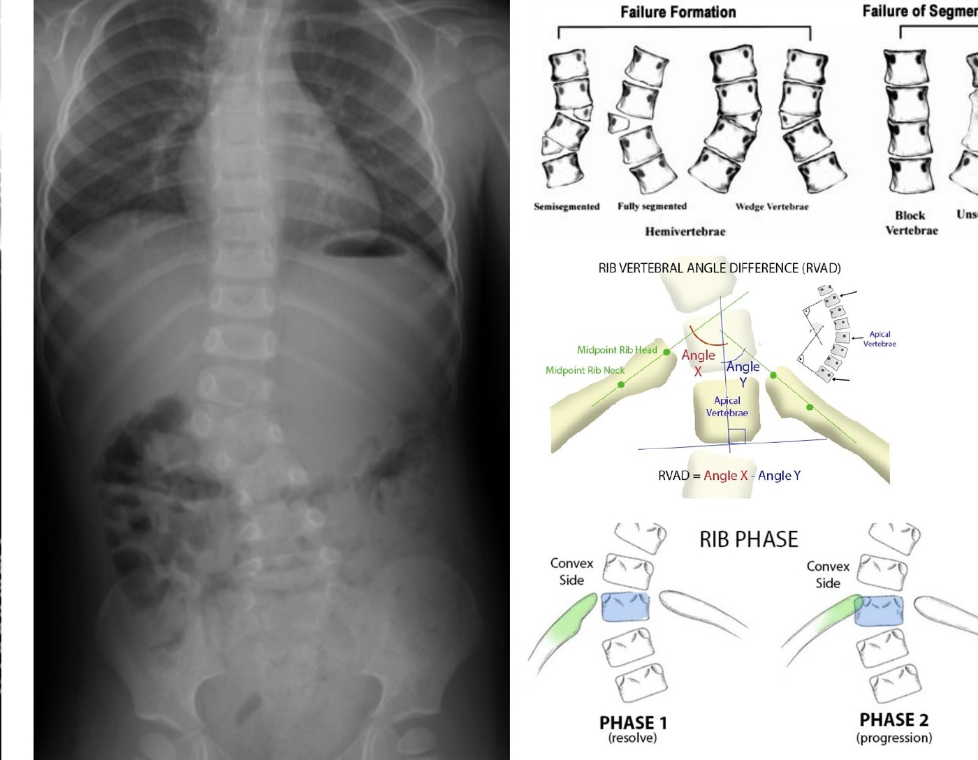
Acute curve at TLJ convex to right side
•One pedicle less over left side, seems unsegmented
•Suspect congenital scoliosis with hemivertebra
History
Birth history
•Walking age, motor development
•Other associated problems e.g. Klippel Feil, NF, VACTERL
Examination
•Clinically check maturity, Tanner whitehouse
•Shoulder balance
•Rib or loin hump
•Truncal shift or listing
[Note:
•Truncal shift is measured by plotting the central sacral vertical line (CSVL) and measuring the deviation of the midpoint of the two widest points of the rib cage along a horizontal parallel line.
•Listing is measured by plotting the C7 plumb line and measuring the deviation between the mid-point of the intercrestal line.
•Hideki Shigematsu, Jason Cheung et al 2017]
•Neurology
•Gait
•Skin stigmata, café au lait
Investigations
XR
•CT
•MRI for intraspinal abnormalities
Internal organ screening (renal USG, echo)
Management
•Aim – arrest progression. Achieve a balanced spine while preserving as much spinal growth as possible
Natural history according to McMaster’s paper
485 patients 75% need surgical treatment
•Prognosis depends on type of anomaly and the anatomical site
•Progression (overall 75% require OT)
•Type: Mixed > unilateral bar > fully segmented hemivertebrae > partially segmented hemivertebrae > incarcerated hemivertebrae (small vertebra sit on nongrowing vertebra) > nonsegmented hemivertebrae
•Position: TLJ worse
•Age
Treatment:
•Observation for cases where there is absence of progression (incarcerated, non segmental, some partially segmented hemivertebrae)
•Operation
•Early In situ fusion for minimal deformity, short segment fusion
•Young : A+P fusion
•Older: P fusion
•Hemivertebrectomy when there is marked truncal imbalance as it allows for deformity correction, for children younger than 5 but flexible curves <40
•Convex hemiepiphysiodesis for unilateral failure of formation, children younger than 5 with short progressive curves <40-50degrees
•Spinal column shortening resection for rigid, severe deformities that present late and have severe decompensation
•Growing rods maximize growth but they are controversial in managing the curve progression
What is congenital scoliosis?
•Failure to form normal vertebrae at gestation week 4-6 [failure of formation / segmentation / mixed]
McMaster classification
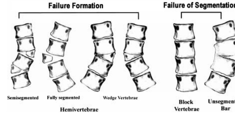
•Failure of formation can be further divided according to
•Segmentation (segmented, semisegmented and non segmented)
•Extent (partial: wedge vs complete: hemivertebrae)
•Most sporadic
Association:
•Extraspinal (VACTERL / KF)
•Intraspinal (Chiari malformation, tethered cord, syrinx)
•Early onset scoliosis
•Infantile idiopathic scoliosis
•More common in boys
•More left sided thoracic curve
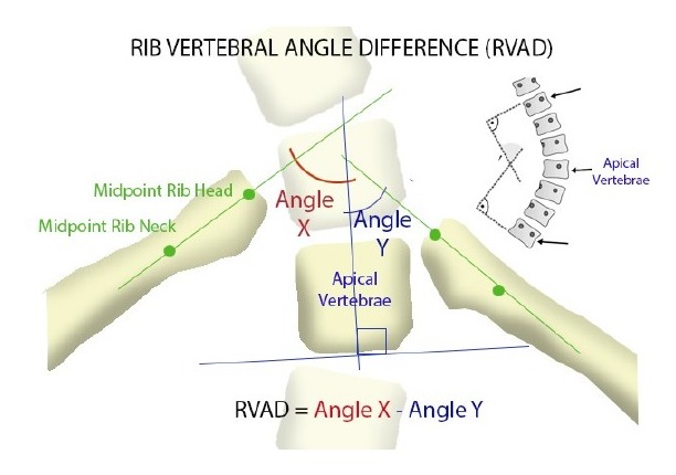
•Look at apical RVAD and phase
•Rib vertebrae angle difference, >20 more progression
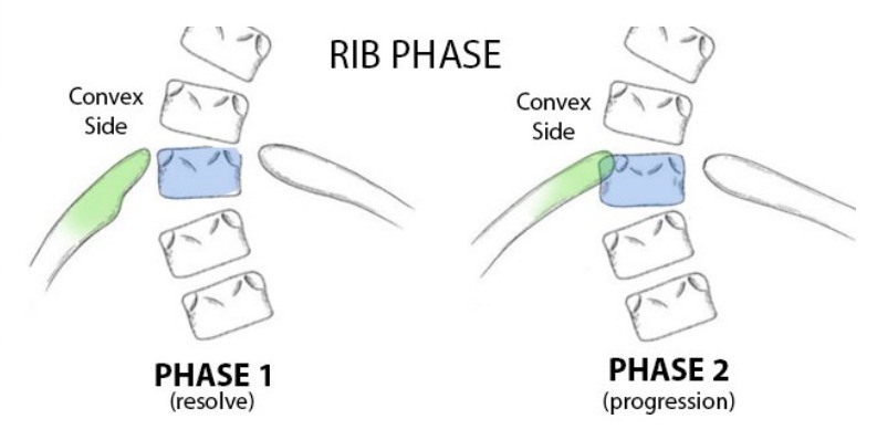
•Phase I (rib not overlap with vertebrae) vs Phase II (rib overlap with apical vertebrae), phase II more progression
•Cobb <30, RVAD<20 – observe
•Cobb >30, RVAD >20, phase II – brace
Cobb >50 – surgery
•Growing rods