Clean FRCS notes with inline figures
•AIS
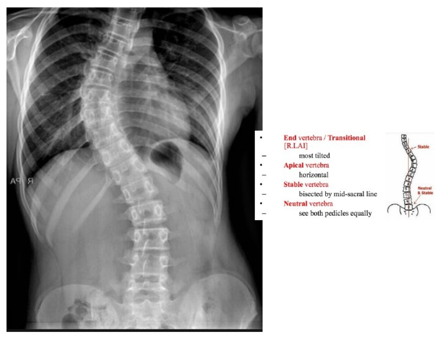
•XR showing scoliosis, with major right thoracic curve. Apex as T8/9. Cobb angle 40 degrees
•Shoulders and pelvis level
•Truncal shift and truncal listing
•No vertebral anomalies such as hemivertebrae or unsegmented bar. Ribs also look normal.
•Risser sign 3. Triradiate cartilage status (0 vs 5 triradiate cartilage not fused at 0)
•Sagittal view to look for thoracic hypokyphosis
•Causes of scoliosis (7)
•Idiopathic
•Postural
•Compensatory (LLD)
•NM (CP)
•Syndromic (NF / marfan / OI / SED…)
•Congenital (Bar / hemivertebrae, eg VACTERL / KF)
•Pathological (Osteoblastoma)
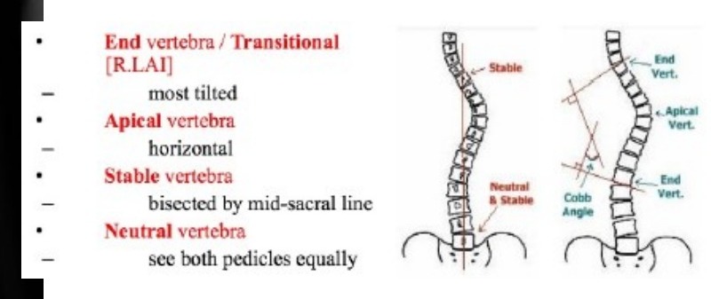
•Lenke classification, King classification, Peking union medical college, Labelle 3D
•Major thoracic curve
History
•1. Birth history
•2. Developmental history
•3. FHx of scoliosis
•4. Current sx
•Age of onset and progression
•Pain or neurological sx
•Visceral problems
•5. Assess maturity: menarche, growth spurt
Further assessment
Confirm diagnosis
•Assess severity
•Estimate risk of progression
•Young presentation: age 9-13, >2y before menarche, growth rate >2cm/yr, risser 0-1, hand XR capped stage (TWIII), open triradiate cartilage
•Severity of curve
•Location of curve (Thoracic spine, double major curve)
Examination
General:
•1. body height, arm span, sitting height
•2. Cutaneous lesions
•3. Ligamentous laxity/ marfanoid features
•4. Spinal dysraphism
Standing:
•Coronal profile: Shoulder and pelvis symmetry, rib or loin hump, decompensation: truncal shift and list
•Sagittal profile: hypokyphosis / kyphoscoliosis
•Bend forward – flexibility
•Gait: scissoring gait
Sit:
•Postural scoliosis
Supine:
•Neuro exam
•Superficial abdominal reflexes
•Finish by checking maturity (secondary sexual characteristics)
Indication for MRI
•Left sided thoracic curve
•Short angular curve
•Angled kyphosis
•Neurology
•Pain
•<10yr + >20 deg
•Rapid curve progression
Management
•Goal is to stop progression and correct deformity, improve truncal balance, improve cosmesis
•Brace
•The aim of the brace is to slow down curve progression, the effect is dosage related ( >20 hr/day → success rate of 93%). Usually will keep bracing till skeletal maturity as curve progression after that will be min. (Weinstein Braist trial NEJM 2013, 72% success rate cw 48% with observation)
•Boston brace if apex below T7
•Milwaukee brace if apex above T7
•Weaning of brace at skeletal maturity (no growth in 18 months, risser 4, menarche >2yr, DR and DU physis closed) in general is done in a gradual manner, no hard science in how it should be weaned. ( e.g. spread over 4/12, stepwise hr/day)
Surgery
Indications for surgery:
•Curves greater than 45 degrees in growing patient or >50 when growth has stopped
Aims of operation:
•Correct deformity and achieve square fusion block with <2cm listing and <20 deg tilt
•Include all structural curves while saving fusions segment
Stable fusion and prevent complications
Key elements of surgery:
Posterior approach
•Subperiosteal dissection till transverse processes
•Identify upper and lower instrumented vertebrae (ASF: end to end. PSF: proximal – end; distal Lenke I-II most distal VB touched by CVSL, III-VI distal end VB)
•Pedicle screws insertion, navigation guided
•If want to correct tilt, can fuse up or down, if want to correct truncal shift, can fuse down
•Reduction techniques: Harrington forces, cantilever bending, translation, rod rotation, direct vertebral rotation
•Insert set screw while holding the rod in place
•Decortication, bone graft to interlaminar space and into facets
•Growing rods if very immature children to prevent crankshaft phenomenon
•Continuous growth of anterior vertebrae while posterior is fused
•Leads to exaggerated lordosis and recurrence of deformity
Old implants used in scoliosis surgery:
•Harrington rod
•Luque rod
•Sublaminar wires
•Difficult to control rotation, as mainly distraction forces
•Risser sign
•I = prepubertal
•II = growth spurt
•III = decrease growth rate
•IV = variable
Disadvantage:
•Peak height velocity and 2/3 of growth spurt occurs before Risser I
•Curve not always stop at Risser IV
the screening program in HK
•Paul: 1995 start by prof luk
•3 tier system (starting in P5)
•FBT + ATR
•Scoliometer 7deg ~20deg cobbs -
•Paul: look for assymetric hump
•Hump convex side
•=/= cobb angle
•>=15, refer scoliosis center
•5-14, Moire topography → >=2 lines → XR → >20 deg → refer specialist hospital
•<5, rerefer screening program
Complications of bracing
•GI regurgitation
•Skin irritation, dermatitis or folliculitis
•Pressure
•Self esteem
Why surgery
•Natural history as outlined by Weinstein’s paper in the 1980s and early 2000s)
•Progression of curve (>50 deg → more likely SOB, >70 deg → decreased lung function, >100 deg increase risk of death)
•Increase in mortality
•Cardiopulmonary compromise
•Truncal imbalance
•Back pain
•Compare and contrast A or P fusion
•Anterior
•Adv: short segment, increase correction power by discectomy, for hypokyphosis (allow anterior column shortening)
•Disadv: difficult exposure
•Posterior
•Adv: Easy exposure
•Disadv: long segment, difficult full correction
Combined: indications
Stiff curve (>50 deg residual on FB XR)
•Severe curve (>70)
•Immature (female <10, male <13) to prevent crankshaft