FRCS Paediatric Ortho — improved notes
FRCS Paediatric Ortho — visual inline notes

118. Supracondylar fracture

visual-inline rebuild1 extracted figuresspeaker notes XML73 study lines6 Q&A

Individual slide images have been cropped from the rendered slide and inserted beside the matching note section. The full slide remains below only for layout/audit.

Clean FRCS notes with inline figures

Supracondylar fracture

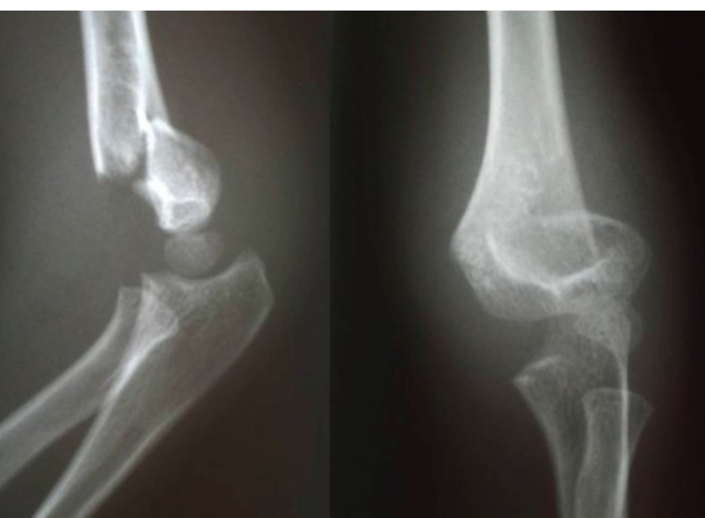
Radiographic description

•Age by CRMTOL (around 2-3 y.o.)
•Pattern of fracture (extension, flexion, Gartland → 1 minimal deformity, 2 anterior cortex broken, posterior cortex intact, 3 posterior cortex not intact)
•Direction of displacement

Gartland classification: type III

•ATLS

Ample history

•r/o fracture in same limb (esp distal radius)

Examination
Clinical photograph
Fig 1. Clinical photographtop left
•Local: Wound, pucker sign, neurological deficit (overall most common is AIN. PL – median, PM – radial, flexion - ulnar), vascular status (radial pulse CR), compartment
•[Nerve inj [OB]: AIN then radial; ulnar most common in flexion type. nearly all cases of neurapraxia following SCH fractures resolve spontaneously → further diagnostic studies are not indicated in the acute setting]
•Systemic: HI, secondary survey
•If any vascular, try emergent CR under conscious sedation, need definitive K wire fixation
•Aim of surgery is restore the alignment in all three planes to prevent late complications such as cubital varus deformity

Most common scenario:

•Pulseless, CR <2sec (Perfusion of UL is well) [I would inform the vascular surgeon on call to be on standby in case the hand perfusion becomes worse after reduction]
•Consent parent for CR +/- OR + KFW as emergent OT (pulseless, pale), urgent – within same day, not to wait overnight (pulseless, pink) JBJS 2015 Skaggs
•In the theatre, the set up of the II and the help of a good assistant is key
•Supine with arm board, II from the end of the bed parallel to the bed

CR maneuver:

•Traction in line with humerus in 20 degrees elbow flexion +/- milking maneuver +/- hyperextension to disengage fracture
•Correct valgus/ varus and rotation by rotating forearm (Supination to relax BR in PL displacement, pronation to close up fracture gap for PM displacement)
•Direct pressure on distal fragment to correct translational deformity
•Thumb over olecranon to reduce extension deformity while maintaining traction
•Assessment of reduction: clinically if hand can touch the shoulder, XR → (need AP, lateral, IO and EO views)
•Anterior humeral line cutting middle 1/3 of capitellum
•Baumann’s angle (angle between long axis of humerus and proximal edge of lateral condyle epiphysis, compare with normal side <5deg difference, N 75 to 80)
•Oblique views to look at the respective columns

Pinning:

•According to AAOS guideline, will insert 2 divergent 1.6mm lateral pins, with maximum spread at fracture site, bicortical fixation, and avoid cross at fracture site
•If still unstable will add 1 more lateral pin / medial pin with mini open wound, dissection down bone, insert K Wire under direct vision with elbow in extension (Larson showed that 3 divergent lateral pins are at least as stable as standard crossed pins JPO 2006)
•(If develop ulnar nerve palsy: likely neuropraxia, closely observe, most return by 3 weeks)
•Cut and bend k wires
•Cast
•Postop monitor perfusion of hand, neurology and also pain (compartment syndrome) for 24-48hrs (Robb JBJS 2009)
•After pinning, wait for circulation to return
•- Extend elbow and wait 15mins after pinning before deciding on OR

Indication of emergency OR:

•Still white
•Pink but pulseless + no anatomical reduction
•Pre CR pulse+, post CR pulse-
•- if hand remains perfused, then slab the arm in 40-60deg of flexion to avoid further vascular compromise
•For nerve injury
•CR for all

Post CR:

•Anatomical reduction → observe
•Suboptimal → explore (check if nerve jamming within fracture site)

Open reduction:

•Lazy S incision, proximal medial to bicep, distal between PT and BR→ watch out for lateral cutaneous nerve of forearm (2cm lateral to bicep tendon) → release bicipital aponeurosis (lat → med, AVN), full supination to protect PIN → retract biceps and brachialis medially and BR laterally
•Post reduction monitor overnight, Q1H charting
•Inside out wound, gustilo 1 in paediatric cases: debridement/ Abx not necessary
•Pink pulseless even after pinning – can be managed with observation
•Most actually still have brachial artery thrombosis in pink pulseless, but no long term sequalae due to adequate collaterals
•(Carbonell et al EPOSNA annual meeting 2017) – no long term sequelae at 20yrs

Postop developed pulseless hand, what to do?

•Complete the rest of vascular examination, assess CR, colour and warmth

Assess intraop XR, reduction, any gap?

•If reduction perfect, pink pulseless, may observe as it may be from vascular spasm
•If reduction not perfect, or white pulseless hand, release backslab and extend arm. Still no improvement, contact vascular team for urgent exploration +/- repair

How to avoid ulnar nerve palsy when medial pinning?

•Extend elbow
•Direct visualization
•Oscillating mode
•Protect with drill sleeve

Indications for medial pinning? (Skaggs JBJS 2001)

•Unstable after 2 lateral pins – relative
•Medial comminution
•Fracture pattern (proximal medial to distal lateral)

Indications for exploration of AIN

•Postop (iatrogenic)
•Fracture site gap
•Open fracture

Direct Q&A

Q1 What are the radiographic findings for Supracondylar fracture?
•Age by CRMTOL (around 2-3 y.o.)
•Pattern of fracture (extension, flexion, Gartland → 1 minimal deformity, 2 anterior cortex broken, posterior cortex intact, 3 posterior cortex not intact)
•Direction of displacement
Q2 What examination findings should be assessed in Supracondylar fracture?
•Local: Wound, pucker sign, neurological deficit (overall most common is AIN. PL – median, PM – radial, flexion - ulnar), vascular status (radial pulse CR), compartment
•[Nerve inj [OB]: AIN then radial; ulnar most common in flexion type. nearly all cases of neurapraxia following SCH fractures resolve spontaneously → further diagnostic studies are not indicated in the acute setting]
•Systemic: HI, secondary survey
•If any vascular, try emergent CR under conscious sedation, need definitive K wire fixation
•Aim of surgery is restore the alignment in all three planes to prevent late complications such as cubital varus deformity
Q3 What are the indications listed for Supracondylar fracture?
•Still white
•Pink but pulseless + no anatomical reduction
•Pre CR pulse+, post CR pulse-
•- if hand remains perfused, then slab the arm in 40-60deg of flexion to avoid further vascular compromise
•For nerve injury
•CR for all
Q4 Postop developed pulseless hand, what to do?
•Complete the rest of vascular examination, assess CR, colour and warmth
Q5 Assess intraop XR, reduction, any gap?
•If reduction perfect, pink pulseless, may observe as it may be from vascular spasm
•If reduction not perfect, or white pulseless hand, release backslab and extend arm. Still no improvement, contact vascular team for urgent exploration +/- repair
Q6 How to avoid ulnar nerve palsy when medial pinning?

Extend elbow

•Direct visualization
•Oscillating mode
•Protect with drill sleeve

Raw speaker-note hierarchy audit

#lvlmarLindentlogicaloriginal note line
10NoneNone0Supracondylar fracture
20NoneNone0Describe Xray:
30171450-1714501Age by CRMTOL (around 2-3 y.o.)
40171450-1714501Pattern of fracture (extension, flexion, Gartland → 1 minimal deformity, 2 anterior cortex broken, posterior cortex intact, 3 posterior cortex not intact)
50171450-1714501Direction of displacement
60000Gartland classification: type III
70000ATLS
80000Ample history
90000r/o fracture in same limb (esp distal radius)
100000PE:
110000Local: Wound, pucker sign, neurological deficit (overall most common is AIN. PL – median, PM – radial, flexion - ulnar), vascular status (radial pulse CR), compartment
120000[Nerve inj [OB]: AIN then radial; ulnar most common in flexion type. nearly all cases of neurapraxia following SCH fractures resolve spontaneously → further diagnostic studies are not indicated in the acute setting]
130000Systemic: HI, secondary survey
140000If any vascular, try emergent CR under conscious sedation, need definitive K wire fixation
150000Aim of surgery is restore the alignment in all three planes to prevent late complications such as cubital varus deformity
160000Most common scenario:
170171450-1714501Pulseless, CR <2sec (Perfusion of UL is well) [I would inform the vascular surgeon on call to be on standby in case the hand perfusion becomes worse after reduction]
180171450-1714501Consent parent for CR +/- OR + KFW as emergent OT (pulseless, pale), urgent – within same day, not to wait overnight (pulseless, pink) JBJS 2015 Skaggs
190000In the theatre, the set up of the II and the help of a good assistant is key
200000Supine with arm board, II from the end of the bed parallel to the bed
210000CR maneuver:
220171450-1714501Traction in line with humerus in 20 degrees elbow flexion +/- milking maneuver +/- hyperextension to disengage fracture
230171450-1714501Correct valgus/ varus and rotation by rotating forearm (Supination to relax BR in PL displacement, pronation to close up fracture gap for PM displacement)
240171450-1714501Direct pressure on distal fragment to correct translational deformity
250171450-1714501Thumb over olecranon to reduce extension deformity while maintaining traction
260000Assessment of reduction: clinically if hand can touch the shoulder, XR → (need AP, lateral, IO and EO views)
271628650-1714502Anterior humeral line cutting middle 1/3 of capitellum
281628650-1714502Baumann’s angle (angle between long axis of humerus and proximal edge of lateral condyle epiphysis, compare with normal side <5deg difference, N 75 to 80)
291628650-1714502Oblique views to look at the respective columns
300000Pinning:
310171450-1714501According to AAOS guideline, will insert 2 divergent 1.6mm lateral pins, with maximum spread at fracture site, bicortical fixation, and avoid cross at fracture site
320171450-1714501If still unstable will add 1 more lateral pin / medial pin with mini open wound, dissection down bone, insert K Wire under direct vision with elbow in extension (Larson showed that 3 divergent lateral pins are at least as stable as standard crossed pins JPO 2006)
332002(If develop ulnar nerve palsy: likely neuropraxia, closely observe, most return by 3 weeks)
340171450-1714501Cut and bend k wires
350171450-1714501Cast
360171450-1714501Postop monitor perfusion of hand, neurology and also pain (compartment syndrome) for 24-48hrs (Robb JBJS 2009)
370000After pinning, wait for circulation to return
380000- Extend elbow and wait 15mins after pinning before deciding on OR
390171450-1714501Indication of emergency OR:
401628650-1714502Still white
411628650-1714502Pink but pulseless + no anatomical reduction
421628650-1714502Pre CR pulse+, post CR pulse-
430000- if hand remains perfused, then slab the arm in 40-60deg of flexion to avoid further vascular compromise
440000For nerve injury
450171450-1714501CR for all
460171450-1714501Post CR:
471628650-1714502Anatomical reduction → observe
481628650-1714502Suboptimal → explore (check if nerve jamming within fracture site)
490000Open reduction:
500000Lazy S incision, proximal medial to bicep, distal between PT and BR→ watch out for lateral cutaneous nerve of forearm (2cm lateral to bicep tendon) → release bicipital aponeurosis (lat → med, AVN), full supination to protect PIN → retract biceps and brachialis medially and BR laterally
510000Post reduction monitor overnight, Q1H charting
520000Inside out wound, gustilo 1 in paediatric cases: debridement/ Abx not necessary
530000Pink pulseless even after pinning – can be managed with observation
540000Most actually still have brachial artery thrombosis in pink pulseless, but no long term sequalae due to adequate collaterals
550000(Carbonell et al EPOSNA annual meeting 2017) – no long term sequelae at 20yrs
560000Postop developed pulseless hand, what to do?
570228600-2286001Complete the rest of vascular examination, assess CR, colour and warmth
580228600-2286001Assess intraop XR, reduction, any gap?
590228600-2286001If reduction perfect, pink pulseless, may observe as it may be from vascular spasm
600228600-2286001If reduction not perfect, or white pulseless hand, release backslab and extend arm. Still no improvement, contact vascular team for urgent exploration +/- repair
610NoneNone0How to avoid ulnar nerve palsy when medial pinning?
620171450-1714501Extend elbow
630171450-1714501Direct visualization
640171450-1714501Oscillating mode
650171450-1714501Protect with drill sleeve
660000Indications for medial pinning? (Skaggs JBJS 2001)
670171450-1714501Unstable after 2 lateral pins – relative
680171450-1714501Medial comminution
690171450-1714501Fracture pattern (proximal medial to distal lateral)
700000Indications for exploration of AIN
710171450-1714501Postop (iatrogenic)
720171450-1714501Fracture site gap
730171450-1714501Open fracture

Improved semantic hierarchy used for the study notes

#semantic levelsection?rewritten line
10Supracondylar fracture
20yesRadiographic description
31Age by CRMTOL (around 2-3 y.o.)
41Pattern of fracture (extension, flexion, Gartland → 1 minimal deformity, 2 anterior cortex broken, posterior cortex intact, 3 posterior cortex not intact)
51Direction of displacement
60yesGartland classification: type III
71ATLS
80yesAmple history
91r/o fracture in same limb (esp distal radius)
100yesExamination
111Local: Wound, pucker sign, neurological deficit (overall most common is AIN. PL – median, PM – radial, flexion - ulnar), vascular status (radial pulse CR), compartment
121[Nerve inj [OB]: AIN then radial; ulnar most common in flexion type. nearly all cases of neurapraxia following SCH fractures resolve spontaneously → further diagnostic studies are not indicated in the acute setting]
131Systemic: HI, secondary survey
141If any vascular, try emergent CR under conscious sedation, need definitive K wire fixation
151Aim of surgery is restore the alignment in all three planes to prevent late complications such as cubital varus deformity
160yesMost common scenario:
171Pulseless, CR <2sec (Perfusion of UL is well) [I would inform the vascular surgeon on call to be on standby in case the hand perfusion becomes worse after reduction]
181Consent parent for CR +/- OR + KFW as emergent OT (pulseless, pale), urgent – within same day, not to wait overnight (pulseless, pink) JBJS 2015 Skaggs
191In the theatre, the set up of the II and the help of a good assistant is key
201Supine with arm board, II from the end of the bed parallel to the bed
210yesCR maneuver:
221Traction in line with humerus in 20 degrees elbow flexion +/- milking maneuver +/- hyperextension to disengage fracture
231Correct valgus/ varus and rotation by rotating forearm (Supination to relax BR in PL displacement, pronation to close up fracture gap for PM displacement)
241Direct pressure on distal fragment to correct translational deformity
251Thumb over olecranon to reduce extension deformity while maintaining traction
261Assessment of reduction: clinically if hand can touch the shoulder, XR → (need AP, lateral, IO and EO views)
272Anterior humeral line cutting middle 1/3 of capitellum
282Baumann’s angle (angle between long axis of humerus and proximal edge of lateral condyle epiphysis, compare with normal side <5deg difference, N 75 to 80)
292Oblique views to look at the respective columns
300yesPinning:
311According to AAOS guideline, will insert 2 divergent 1.6mm lateral pins, with maximum spread at fracture site, bicortical fixation, and avoid cross at fracture site
321If still unstable will add 1 more lateral pin / medial pin with mini open wound, dissection down bone, insert K Wire under direct vision with elbow in extension (Larson showed that 3 divergent lateral pins are at least as stable as standard crossed pins JPO 2006)
332(If develop ulnar nerve palsy: likely neuropraxia, closely observe, most return by 3 weeks)
341Cut and bend k wires
351Cast
361Postop monitor perfusion of hand, neurology and also pain (compartment syndrome) for 24-48hrs (Robb JBJS 2009)
371After pinning, wait for circulation to return
381- Extend elbow and wait 15mins after pinning before deciding on OR
390yesIndication of emergency OR:
402Still white
412Pink but pulseless + no anatomical reduction
422Pre CR pulse+, post CR pulse-
431- if hand remains perfused, then slab the arm in 40-60deg of flexion to avoid further vascular compromise
441For nerve injury
451CR for all
460yesPost CR:
472Anatomical reduction → observe
482Suboptimal → explore (check if nerve jamming within fracture site)
490yesOpen reduction:
501Lazy S incision, proximal medial to bicep, distal between PT and BR→ watch out for lateral cutaneous nerve of forearm (2cm lateral to bicep tendon) → release bicipital aponeurosis (lat → med, AVN), full supination to protect PIN → retract biceps and brachialis medially and BR laterally
511Post reduction monitor overnight, Q1H charting
521Inside out wound, gustilo 1 in paediatric cases: debridement/ Abx not necessary
531Pink pulseless even after pinning – can be managed with observation
541Most actually still have brachial artery thrombosis in pink pulseless, but no long term sequalae due to adequate collaterals
551(Carbonell et al EPOSNA annual meeting 2017) – no long term sequelae at 20yrs
560yesPostop developed pulseless hand, what to do?
571Complete the rest of vascular examination, assess CR, colour and warmth
580yesAssess intraop XR, reduction, any gap?
591If reduction perfect, pink pulseless, may observe as it may be from vascular spasm
601If reduction not perfect, or white pulseless hand, release backslab and extend arm. Still no improvement, contact vascular team for urgent exploration +/- repair
610yesHow to avoid ulnar nerve palsy when medial pinning?
621Extend elbow
631Direct visualization
641Oscillating mode
651Protect with drill sleeve
660yesIndications for medial pinning? (Skaggs JBJS 2001)
671Unstable after 2 lateral pins – relative
681Medial comminution
691Fracture pattern (proximal medial to distal lateral)
700yesIndications for exploration of AIN
711Postop (iatrogenic)
721Fracture site gap
731Open fracture