FRCS Paediatric Ortho — improved notes
FRCS Paediatric Ortho — visual inline notes

138. Triplane ankle fracture

visual-inline rebuild1 extracted figuresspeaker notes XML42 study lines2 Q&A

Individual slide images have been cropped from the rendered slide and inserted beside the matching note section. The full slide remains below only for layout/audit.

Clean FRCS notes with inline figures

•AP and lateral Xray of ankle of a skeletally immature patient
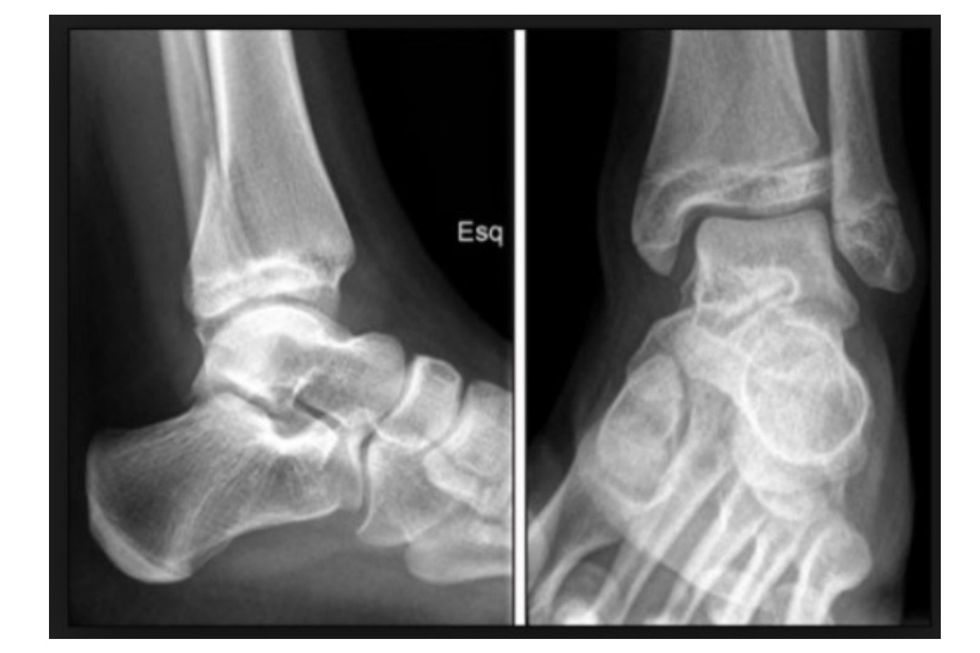
Radiograph / imaging
Fig 1. Radiograph / imagingtop left
•With fracture over distal tibia involving epiphysis
•On AP view it is SH III, on lateral view it is SH II (Thurstan Holland fragment)
•Distal fragment is displaced medial and posterior
•50% associated with fibular fracture
•Triplane fracture (fracture courses through coronal, transverse and sagittal plane)
•Transitional fracture
•This is an intra-articular fracture
•MOI: ER
•Acute: systemically rule out other injuries, locally look for wound, compartment syndrome and document distal NV condition. RICE, slab, analgesics
•Since displacement > 2mm

Definitive: aim at anatomical reduction and stable fixation

•CT
•Lateral (more common) vs medial (judged by distal coronal fragment)
•2 part, 3 part, 4 part
•CR +/- OR, fix with screw (interfragmental compression with screws parallel to physis)
•CR technique: for 2 part - lateral (IR), medial (abduction)
•AL approach, reduce with dorsiflexion + IR +/- pronation, or compression using Weber bone reduction forceps, fixed with 4.0 cannulated screw (transmetaphyseal and transepiphyseal screws)
•Need OT for 3-4 part (sequence: posterior fragment → fibula → AL fragment)
•Advise for removal of transepiphyseal screws. Cadaveric studies (JPO 2005 Charlton) have shown significant increase in peak intraarticular contact pressures over baseline

•Distal tibia physis close at 16yo, distal fibula closes 1 year after
•Contributed to 40% of tibia growth and 20% of LL growth
•All ligaments attach distal to physis
•Distal tibial physis fuse
•Age 11-13 in boys
•Transitional fracture ~ 18 months
•Coronal: cross epiphysis
•Axial: split physis
•Sagittal: exit in metaphysis
•Can be 2, 3, 4 parts

2 parts (named after coronal distal fragment):

•Lateral triplane: supination-external rotation (most common)
•Medial triplane: adduction

3 parts (separate Tillaux fragment):

•SH 3 on AP
•4 part: (Tillaux + AM fragment)
•50% associated with fibular fracture
•Medial os subtibile 20%
•Lateral os subfibulare 1%
•Sequence of distal tibia physis closure: central → anteromedial → posteromedial → anterolateral
•Kump’s bump [radiopedia: superior convexity of the growth plate located in the anteromedial quadrant of the distal tibia physis, which is the first site of physeal fusion. Closure usually occurs at about 12-13 years of age.

Direct Q&A

Q1 What diagnostic or radiographic findings support Triplane ankle fracture?
•AP and lateral Xray of ankle of a skeletally immature patient
•With fracture over distal tibia involving epiphysis
•On AP view it is SH III, on lateral view it is SH II (Thurstan Holland fragment)
•Distal fragment is displaced medial and posterior
•50% associated with fibular fracture
•Triplane fracture (fracture courses through coronal, transverse and sagittal plane)
•Transitional fracture
•This is an intra-articular fracture
•MOI: ER
•Acute: systemically rule out other injuries, locally look for wound, compartment syndrome and document distal NV condition. RICE, slab, analgesics
Q2 What is the definitive management for Triplane ankle fracture?

CT

•Lateral (more common) vs medial (judged by distal coronal fragment)
•2 part, 3 part, 4 part
•CR +/- OR, fix with screw (interfragmental compression with screws parallel to physis)
•CR technique: for 2 part - lateral (IR), medial (abduction)
•AL approach, reduce with dorsiflexion + IR +/- pronation, or compression using Weber bone reduction forceps, fixed with 4.0 cannulated screw (transmetaphyseal and transepiphyseal screws)
•Need OT for 3-4 part (sequence: posterior fragment → fibula → AL fragment)
•Advise for removal of transepiphyseal screws. Cadaveric studies (JPO 2005 Charlton) have shown significant increase in peak intraarticular contact pressures over baseline

Raw speaker-note hierarchy audit

#lvlmarLindentlogicaloriginal note line
10NoneNone0AP and lateral Xray of ankle of a skeletally immature patient
20NoneNone0With fracture over distal tibia involving epiphysis
30NoneNone0On AP view it is SH III, on lateral view it is SH II (Thurstan Holland fragment)
40NoneNone0Distal fragment is displaced medial and posterior
50NoneNone050% associated with fibular fracture
60NoneNone0Triplane fracture (fracture courses through coronal, transverse and sagittal plane)
70NoneNone0Transitional fracture
80NoneNone0This is an intra-articular fracture
90NoneNone0MOI: ER
100NoneNone0Acute: systemically rule out other injuries, locally look for wound, compartment syndrome and document distal NV condition. RICE, slab, analgesics
110000Since displacement > 2mm
120NoneNone0Definitive: aim at anatomical reduction and stable fixation
130NoneNone0CT
140NoneNone0Lateral (more common) vs medial (judged by distal coronal fragment)
150NoneNone02 part, 3 part, 4 part
160NoneNone0CR +/- OR, fix with screw (interfragmental compression with screws parallel to physis)
170000CR technique: for 2 part - lateral (IR), medial (abduction)
180NoneNone0AL approach, reduce with dorsiflexion + IR +/- pronation, or compression using Weber bone reduction forceps, fixed with 4.0 cannulated screw (transmetaphyseal and transepiphyseal screws)
190NoneNone0Need OT for 3-4 part (sequence: posterior fragment → fibula → AL fragment)
200NoneNone0Advise for removal of transepiphyseal screws. Cadaveric studies (JPO 2005 Charlton) have shown significant increase in peak intraarticular contact pressures over baseline
210NoneNone0=================================
220NoneNone0Distal tibia physis close at 16yo, distal fibula closes 1 year after
230NoneNone0Contributed to 40% of tibia growth and 20% of LL growth
240NoneNone0All ligaments attach distal to physis
250NoneNone0Distal tibial physis fuse
260NoneNone0Age 11-13 in boys
270NoneNone0Transitional fracture ~ 18 months
280NoneNone0Coronal: cross epiphysis
290NoneNone0Axial: split physis
300NoneNone0Sagittal: exit in metaphysis
310NoneNone0Can be 2, 3, 4 parts
320NoneNone02 parts (named after coronal distal fragment):
331NoneNone1Lateral triplane: supination-external rotation (most common)
341NoneNone1Medial triplane: adduction
350NoneNone03 parts (separate Tillaux fragment):
360NoneNone0SH 3 on AP
370NoneNone04 part: (Tillaux + AM fragment)
380NoneNone050% associated with fibular fracture
390NoneNone0Medial os subtibile 20%
400NoneNone0Lateral os subfibulare 1%
410NoneNone0Sequence of distal tibia physis closure: central → anteromedial → posteromedial → anterolateral
420NoneNone0Kump’s bump [radiopedia: superior convexity of the growth plate located in the anteromedial quadrant of the distal tibia physis, which is the first site of physeal fusion. Closure usually occurs at about 12-13 years of age.

Improved semantic hierarchy used for the study notes

#semantic levelsection?rewritten line
10AP and lateral Xray of ankle of a skeletally immature patient
21With fracture over distal tibia involving epiphysis
31On AP view it is SH III, on lateral view it is SH II (Thurstan Holland fragment)
41Distal fragment is displaced medial and posterior
5150% associated with fibular fracture
61Triplane fracture (fracture courses through coronal, transverse and sagittal plane)
71Transitional fracture
81This is an intra-articular fracture
91MOI: ER
101Acute: systemically rule out other injuries, locally look for wound, compartment syndrome and document distal NV condition. RICE, slab, analgesics
111Since displacement > 2mm
120yesDefinitive: aim at anatomical reduction and stable fixation
131CT
141Lateral (more common) vs medial (judged by distal coronal fragment)
1512 part, 3 part, 4 part
161CR +/- OR, fix with screw (interfragmental compression with screws parallel to physis)
171CR technique: for 2 part - lateral (IR), medial (abduction)
181AL approach, reduce with dorsiflexion + IR +/- pronation, or compression using Weber bone reduction forceps, fixed with 4.0 cannulated screw (transmetaphyseal and transepiphyseal screws)
191Need OT for 3-4 part (sequence: posterior fragment → fibula → AL fragment)
201Advise for removal of transepiphyseal screws. Cadaveric studies (JPO 2005 Charlton) have shown significant increase in peak intraarticular contact pressures over baseline
210——
221Distal tibia physis close at 16yo, distal fibula closes 1 year after
231Contributed to 40% of tibia growth and 20% of LL growth
241All ligaments attach distal to physis
251Distal tibial physis fuse
261Age 11-13 in boys
271Transitional fracture ~ 18 months
281Coronal: cross epiphysis
291Axial: split physis
301Sagittal: exit in metaphysis
311Can be 2, 3, 4 parts
320yes2 parts (named after coronal distal fragment):
331Lateral triplane: supination-external rotation (most common)
341Medial triplane: adduction
350yes3 parts (separate Tillaux fragment):
361SH 3 on AP
3714 part: (Tillaux + AM fragment)
38150% associated with fibular fracture
391Medial os subtibile 20%
401Lateral os subfibulare 1%
411Sequence of distal tibia physis closure: central → anteromedial → posteromedial → anterolateral
421Kump’s bump [radiopedia: superior convexity of the growth plate located in the anteromedial quadrant of the distal tibia physis, which is the first site of physeal fusion. Closure usually occurs at about 12-13 years of age.